Syncope
A 3D transesophageal echocardiogram (TEE) (Video 19-1) and M-mode (Fig. 19-1) are shown for a 37-year-old woman with syncope.
QUESTION 1. The abnormality shown is:
A. Aortic stenosis
B. Left atrial myxoma
C. Infective endocarditis
D. Mitral valve prolapse
E. None of the options
View Answer
ANSWER 1: B. The 3D TEE video shows the mitral valve from the left ventricular perspective. The myxoma enters the left ventricle during diastole.
In the M-mode image (Fig. 19-2), the mitral valve (A) is seen (arrow), and the myxoma (B) prolapses into the mitral orifice with each diastole.
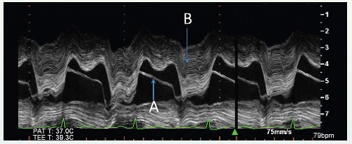 Figure 19-2.
Stay updated, free articles. Join our Telegram channel
Full access? Get Clinical Tree
 Get Clinical Tree app for offline access

|