Figure 4.1
Presentations that may be categorised as fits, faints (unconsciousness) or funny turns, including collapse query cause (transient collapse). True transient loss of consciousness differential diagnosis is represented in the central white box
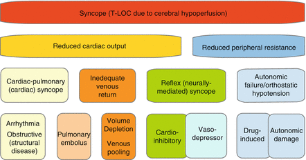
True syncope has a number of potential underlying causes, detailed in Fig. 4.2 and divided by mechanism. It is important to have an accurate early clinical impression of any presentation, in order to avoid inappropriate investigation or reassurance of individuals.


Figure 4.2
Potential underlying causes of cardiac syncope, grouped vertically by physiological mechanism for cerebral hypoperfusion
Approaching the Patient with Syncope
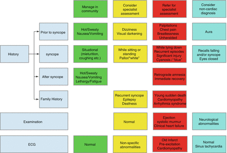
A summary of evaluation findings and indicators of necessity for specialist evaluation following syncope are given in Fig. 4.3. Each aspect of the initial evaluation is detailed below.


Figure 4.3
Suggested clinical indicators of necessity for specialist cardiac referral following presentation with syncope
History
Differentiation between causes of T-LOC is predominantly achieved by careful history-taking, with up to 95 % correctly diagnosed on symptoms alone. Patients may use a variety of terms including “blackout” and “faint” to describe an episode of T-LOC. However, the clinician is often hindered by the inability of patients to describe events accurately; witness observations can equally be unreliable or absent in many cases.
Amnesia surrounding the event itself indicates a high likelihood of T-LOC rather than fall, although bear in mind that elderly patients or those with a head injury may be unaware of the extent of unconsciousness due to retrograde amnesia. Hence, individuals initially assessed under the catch-all “collapse query cause” may require careful consideration, since therapeutic interventions aimed at minimising recurrent syncopal risk will be ineffective against falls due to mechanical and ambulatory factors.
Despite the limitations of patient recollection, it is important to ask specifically about events immediately preceding and after any T-LOC. Moreover, you should attempt to elicit a personal and witness description of the period of unconsciousness to facilitate diagnosis. Personal and family medical history can also be useful in risk-stratification.
Circumstances of Syncope
Reflex, or neurally-mediated syncope, the most common cause in adults, is particularly amenable to diagnosis on history alone. For example: fear, pain, emotional distress, clinical instrumentation or prolonged standing are frequent precipitants. Syncope while supine is very rarely reflex in origin, except when precipitated by such an emotional stimulus. Prodromes of reflex syncope include symptoms of increased vagal tone such as nausea, vomiting, sweating and general fatigue. Situational syncope, which shares an autonomic aetiology, occurs with coughing, swallowing, micturition or defecation; these precipitants are thought to cause increased intrathoracic pressure and subsequent vagal stimulation. Syncope related to shaving in men, or during looking upwards or turning to cross the street may indicate carotid sinus hypersensitivity, another form of neurally-mediated syncope.
Syncope during active exercise is rarely reflex in origin, although the typical increase in parasympathetic tone immediately post-exercise can be responsible for syncope. This post-exertional effect can unmask orthostatic hypotension, where individuals with true autonomic failure are unable to mount a blood pressure response to exercise, but do suffer from the physiological drop in recovery after exertion. However, post-exertional hypotension and bradycardia are common and normal findings in trained athletes who, at onset of exertion, demonstrate rapid increases in heart rate and blood pressure, with similar precipitous falls on resting afterward. Orthostatic hypotension is precipitated by standing, though the drop in blood pressure and resultant syncope may be delayed by several minutes after posture change. While autonomic in origin, orthostatic hypotension is due to disease of the autonomic system rather than an inappropriate reflex in health. Therefore, the prodrome of reflex syncope is absent. A rare variant of autonomic failure can present with post-prandial syncope, particularly with high carbohydrate loads. Dehydration and hot environment (due to skin vasodilation) increase risk for orthostatic hypotension, though this may also contribute to reflex syncope.
Arrhythmic and other cardiac syncope may occur with little or no warning, referred to as unheralded. Alternatively, sudden onset palpitations, indicative of paroxysmal tachyarrhythmia may precede the event. Syncope during exercise or exertion, but excluding early post-exercise syncope, is highly suggestive of a primary cardiac cause.
Epilepsy, an important differential for T-LOC, may be preceded by a stereotypical aura, often specific to the individual. These may include a rising epigastric sensation, distinctive smell or taste, deja vu, or visual aura (but not generally visual field loss or blurring, which are more likely to indicate cardiovascular syncope). Epilepsy may be precipitated by flashing lights, such as during video-game playing and is also seen during sleep deprivation.
Period of Loss of Consciousness
During the period of unconsciousness, the patient should have no recollection of events. If he or she can recall events from the period of unresponsiveness, psychogenic (or non-epileptic) attacks are the most likely diagnosis. Unresponsiveness lasting a long time (more than 2 or 3 min) also suggests a psychogenic cause; this is often associated with waxing and waning of prominent limb and pelvic thrusting. Another useful indicator of psychogenic cause is that the patient’s eyes are generally closed during the episode, unlike syncope and epilepsy, where the eyes remain open. Moreover, about 10 % of psychogenic blackouts initially present with mimicry of status epilepticus.
Epileptic attacks vary in appearance, however, depending on the type of seizure. Generalised seizures are often seen as initial rigidity and fixed extension of limbs, with subsequent symmetrical tonic-clonic limb movements of large amplitude, which gradually decrease in amplitude over the course of the seizure. Importantly, limb movements may commence prior to loss of consciousness, which is not seen in syncope. They very rarely last longer than 1 min, although panicked bystanders may overestimate the duration when reporting collateral history. Biting of the lateral tongue border (rather than tip) and frothing mouth are other signs of generalised epilepsy. Facial cyanosis may also be seen, although cyanosis can equally indicate malignant arrhythmia. Complex partial seizures may present with asymmetric or unilateral twitching, head-turning or lip-smacking; altered consciousness may be present at onset, or become apparent later during the attack. Secondary generalisation, with features akin to generalised epilepsy, may be witnessed.
Limb jerking and other mimics of seizures can occur with syncope, due to secondary cerebral hypoxia. It is important to appreciate this so as to avoid the pitfall of diagnosing a primary seizure in these cases. In fact, they are associated with convulsive syncope of any aetiology, including reflex syncope. Extreme facial pallor, described by witnesses as, “white as a sheet,” while frightening to bystanders, is also indicative of the central vasodilation and preferential vascular flow away from the skin of reflex syncope. In contrast, individuals who experience arrhythmic syncope may be cyanotic, though this appearance is also seen in epileptic seizures. Syncopal loss of consciousness is less than 30 s in duration and generally less than 15 s.
Return of Consciousness
Post-event recovery following epileptic seizures can take over 5 min, with prolonged confusion and disorientation; other features include headache and muscle ache, though bruising due to a fall may confound this symptom. Recovery from reflex syncope is more rapid, although similar autonomic post-event symptoms to the prodrome are frequent, particularly prolonged fatigue and lethargy. In the absence of physical injury, resumption of normal activity following arrhythmic syncope is generally instantaneous, such that patients may not recognise the seriousness of the event. Other cardiac causes of syncope may leave individuals with associated residual symptoms such as breathlessness, chest pain or palpitations.
Past Medical and Medication History
Syncope that occurs in the context of structural or functional heart disease is most likely to be secondary to malignant arrhythmia. Loss of consciousness in individuals with alcoholism, autism, cerebral palsy, learning difficulties or brain injury tends to associate with epilepsy. Primary nervous system disease such as multi-system atrophy and systemic diseases with neuropathic complications, such as diabetic and amyloid neuropathy suggest orthostatic hypotension; this may be exacerbated by medications such as antihypertensives, antidepressants and diuretics. However, antidepressants may also result in cardiac depolarisation and repolarisation anomalies which predispose to malignant arrhythmias, so this should always be considered. Other medications, alcohol and drugs of abuse are also know to affect cardiac electrophysiology adversely, especially in overdose.
Family History
Family history may provide an important marker for more malignant syncope. Close blood relatives of individuals with inherited primary arrhythmia syndromes (e.g. long QT syndrome, Brugada syndrome and catecholaminergic polymorphic tachycardia) or inherited myocardial disorders (e.g. hypertrophic cardiomyopathy, familial dilated cardiomyopathy, arrhythmogenic right ventricular cardiomyopathy) who present with transient loss of consciousness should be thoroughly investigated for these conditions, since the risk of sudden death is high. Similarly, a family history of premature and/or unexplained sudden death may represent undiagnosed cardiovascular genetic conditions in up to half of all cases and should prompt comprehensive cardiac evaluation for inherited disease. A family history of recurrent seizures or epilepsy warrants close evaluation for both familial epilepsy and inherited cardiac conditions, due to the possibility of misdiagnosis of syncope as epilepsy.
Examination
Clinical examination can suggest specific cardiac or neurological causes for syncope, though the vast majority of patients have normal findings especially in reflex syncope.
Obstruction to left ventricular oUTFlow, caused either by hypertrophic cardiomyopathy or aortic stenosis, may be diagnosed clinically by the character of an ejection systolic murmur, in addition to the changes with squatting and/or amplitude of the second heart sound. Similarly atrial myxoma may be suggested by a tumour plop or mitral valve murmur. Signs of heart failure are less specific, though do indicate a likely cardiac cause. Significant difference in bilateral arm blood pressures may suggest aortic pathology, though it may indicate a possible subclavian steal in syncopal patients.
Any focal neurological deficits are strongly suggestive of epilepsy. However, limb weakness, ataxia and cranial nerve dysfunction may indicate vertebrobasilar transient ischaemic attacks (TIA). In general, TIA should not be considered in the differential for syncope in the absence of a coinciding suggestive neurological deficit.
< div class='tao-gold-member'>
Only gold members can continue reading. Log In or Register to continue
Stay updated, free articles. Join our Telegram channel

Full access? Get Clinical Tree


