Surgical Anatomy
Mohamed A. Abdalla1
Bruce Bollen2
Carlos Duran2
Robert M. Savage2
1OUTLINE AUTHOR
2ORIGINAL CHAPTER AUTHORS
▪ KEY POINTS
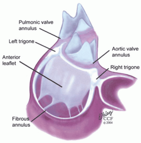
The fibrous skeleton of the heart is formed by the U-shaped cords of the aortic annulus forming the right and left trigones. From the left and right trigones, a fibrous tissue continuum extends around the left and right atrioventricular orifices, forming the annuli fibrosi of the mitral and tricuspid annuli. The mitral annuli firbosi thins posteriorly, permitting pathologic annular dilatation and increased tension on the middle scallop of the PMVL.
The Society of Cardiovascular Anesthesiologists and American Society of Echocardiography (ASE) have jointly developed a 16-segment model of the left ventricle (LV) based on the recommendations of the Subcommittee on Quantification of the ASE Standards Committee, which divides the LV into three levels (basal, mid, and apical). The base and mid levels are divided into six segments and the apex into four.
The three leaflets of the tricuspid valve are the anterior, posterior, and septal. The anterior leaflet is usually the largest leaflet and the posterior leaflet the smallest. The valve has three commissures: anteroseptal commissure, anteroposterior commissure, and posteroseptal commissure.
The pulmonic annulus and valve are attached to the base of the aorta by a fibrous extension of the aortic root called the tendon of conus.
The mitral valve apparatus consists of the fibrous skeleton of the heart, the mitral annulus, mitral leaflets, mitral chordae, and the papillary muscle-ventricular wall complex. There are three nomenclatures used to describe the apparatus: anatomic terminology, SCA/ASE (Carpentier) terminology, and Duran terminology.
The aortic root is comprised of the aortic annulus, valve leaflets, sinuses of Valsalva, and sinotubular junction. Pathologic distortion of any of these components may result in aortic-valve dysfunction.
The aortic valve has three coronary cusps (right, left, and noncoronary) with corresponding expanded sinuses inferiorly defined by attachment of the leaflet and the sinotubular junction.
The heart is supplied by three coronary arteries: the left anterior descending (LAD), the circumflex (LCx), and the right coronary artery (RCA). The LAD supplies the anterior two thirds of the interventricular septum, anterolateral
free wall of the LV, and the infranodal conduction system (Bundle of His, right bundle branch, and left anterior fascicle). The LCx supplies the posterior and inferior (7% of patients) wall of the LV and portions of the posterior fascicle of the LBB. It also supplies the posteromedial papillary muscle. The RCA supplies the right ventricle and the inferior LV (in 85% of patients) in addition to the AV node. Myocardial ischemia produces regional wall-motion abnormalities and dysrhythmias that may be predicted on the basis of coronary circulation.
I. SURGICAL ANATOMY OF THE HEART
A. Fibrous skeleton of the heart
Fibrous skeleton formed by U-shaped cords of aortic annulus extensions forming right trigone, left trigone, and fibrous structure from the right aortic coronary cusp to the root of the pulmonary artery.
Skeleton supports the heart within the pericardium (Fig. 4-1).
B. Cardiac ventricles
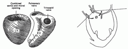
Right ventricle has two openings separated by band of myocardium, crista supraventricularis.
Two openings are tricuspid and pulmonic valves. LV has a common opening at its base shared by aortic root and mitral valve (Fig. 4-2).
Left ventricular outflow tract (LVOT)
defined anteriorly by membranous and muscular portion of interventricular septum
defined posteriorly by anterior leaflet of mitral valve
Ventricle is divided into a 16-segment model.
Divides LV into three levels: basal, mid, and apical. Basal and mid levels are each divided circumferentially into six segments and apical into four (Figs. 4-3, 4-4, 4-5
Stay updated, free articles. Join our Telegram channel

Full access? Get Clinical Tree


