Fig. 19.1
ECG shows a narrow QRS tachycardia
The ECG showed a narrow QRS complex tachycardia. Heart rate was 200 beats per minute (RR 300 ms). The QRS axis is about + 20°. P waves were not clearly visible. ST segment depression was present in I, II, aVF, and V3–V6 leads. ST segment elevation was present in aVR and V1 leads.
What Are the Possible Types of Supraventricular Arrhythmias?
Sinus tachycardia
Reentrant supraventricular tachycardias
Atrioventricular reciprocating tachycardia
Atrioventricular nodal reciprocating tachycardia
Focal atrial tachycardia
Atrial flutter
Atrial fibrillation
The presence of a regular rhythm excludes the hypothesis of atrial fibrillation. There isn’t a clearly visible P wave, probably because it is inside the terminal part of the ventricular complexes. The atrioventricular ratio is 1:1. Heart rate (200 bpm) is too low for a 1:1 atrial flutter, so this hypothesis is unlikely. Even if the morphology of the P wave is not assessable, we can exclude the hypothesis of a sinus tachycardia because there were no physiological causes for having high heart rate at rest. RP interval was shorter than PR and longer than 70 ms. The most likely diagnoses left are therefore atrioventricular reciprocating tachycardia, atrioventricular nodal reciprocating tachycardia, and atrial tachycardia.
Vagal maneuver response may allow to distinguish between the different forms of supraventricular tachycardias. Atrial tachycardia generally is conducted with a transitory high-grade atrioventricular block. Reentrant supraventricular tachycardias end suddenly. In this case, the sinus carotid massage caused a sudden tachycardia interruption (Fig. 19.2).
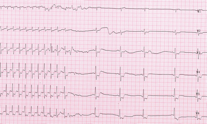
Fig. 19.2
Tachycardia termination during sinus carotid massage
Therefore, the diagnosis was reentrant supraventricular tachycardia. The high heart rate (about 200 bpm) was more suggestive for an atrioventricular reciprocating tachycardia, but this is not a definite clue.
The patient was admitted to the cardiology department to perform an invasive electrophysiological investigation with subsequent catheter ablation of the tachycardia.
Invasive Electrophysiological Investigation (Fig. 19.3)
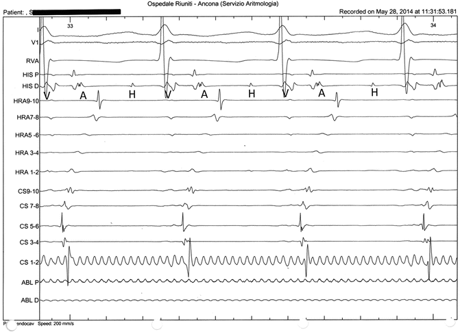
Fig. 19.3
Electrophysiological study. Endocavity ECG during tachycardia. A atrium, H His, V ventricle
[…] eccentric type ventriculo-atrial retrograde conduction. Early retrograde conduction in left posterolateral region (CS 5–6). Induction by programmed atrial pacing of an orthodromic atrioventricular reciprocating tachycardia (concealed accessory pathway). During tachycardia a catheter ablation of the posterolateral accessory pathway with termination of tachycardia was performed. […]
The final diagnosis was atrioventricular reciprocating tachycardia from posterolateral concealed accessory pathway.
What Is the Meaning of ST Segment Depression During Supraventricular Paroxysmal Tachycardia? Can It Be a Sign of Atherosclerotic Coronary Disease?
This patient had a high cardiovascular risk profile (type 2 diabetes mellitus, arterial hypertension, dyslipidemia, overweight, past smoking status), and he was symptomatic for chest pain during tachycardia. Echocardiography and a treadmill test were performed.
Echo: Normal atrial size. Normal left ventricle size and systolic function (EF 60 %). Normal right ventricle size and systolic function (TAPSE 19 mm). Normal diastolic function. Mild mitral regurgitation. Mild tricuspid regurgitation with normal pulmonary arterial pressure. No pericardial effusion.
The exercise test was interrupted at the end of Bruce’s protocol 5th stage. The patient reached the peak exercise with a heart rate of 154 bpm (96 % of theoretical maximum heart rate) and a systolic blood pressure of 170 mmHg (double product: 26,180). Heart rate and blood pressure showed a normal behavior. There were no symptoms of ischemic ECG changes.
We conclude for nonischemic ST alteration. The abnormal repolarization may be the consequence of a distinct pattern of retrograde atrial activation through accessory pathway. This phenomenon (ST depression >2 mm) is more frequent in AV reentrant tachycardia than in AV nodal reentrant tachycardia. Indeed, in orthodromic AV tachycardia, atrial retrograde activation, as assessed by intracardiac electrograms, occurs during the ST segment on the surface ECG. In contrast, in AV nodal reentrant tachycardia, the retrograde atrial activation is most frequently simultaneous with the QRS complex and therefore does not interfere with repolarization [1–4].
19.2 Atrioventricular Nodal Reentrant Tachycardia (AVNRT)
Definition and Epidemiology
Atrioventricular nodal reentrant tachycardia is the most common form of regular paroxysmal supraventricular tachycardia. Although it was classically described more often in the young and women, it can be detected at any age and sex. The overall prevalence of atrioventricular nodal reentrant tachycardia is 2–3 cases per 1000 persons. Atrioventricular nodal reentrant tachycardia is not usually associated with structural heart disease [5–7].
Physiopathology and Classification
The reentrant circuit is confined not only in the compact AV node, but the perinodal atrial tissue has an important role too. Atrioventricular nodal reentrant tachycardia is related to the presence of two functionally and anatomically distinct AV nodal pathways (dual AV nodal pathways). The fast pathway, which conducts more rapidly (PR interval 100–150 ms and AH <220 ms) and has a long refractory period, appears to be located near the apex of Koch’s triangle. The slow pathway, which conducts more slowly (PR interval >220 ms) and has a short refractory period, extends inferoposterior to the compact AV node tissue and extends along the septal margin of the tricuspid annulus at the level of the coronary sinus. The fast pathway constitutes the normal, physiological, AV conduction axis [6].
AVNRT has been categorized into typical or atypical. This categorization is based on the retrograde limb of the circuit: if it is the fast pathway, it is defined typical; if it is the slow pathway, it is atypical. Typical atrioventricular nodal reentrant tachycardia is the more common type (90 %) and involves the slow pathway for antegrade conduction and the fast pathway for retrograde conduction (slow-fast variant). Atypical atrioventricular nodal reentrant tachycardia is less common (10 %) and includes fast-slow and slow-slow variants [6].
An atrial premature complex that is blocked in the fast pathway and is conducted through the slow pathway generally initiates typical AVNRT. If the fast pathway has recovered excitability, the impulse can conduct retrogradely over the fast pathway, resulting in an atrial echo. If the impulse reenters into the slow pathway, typical AVNRT may be initiated. Seldom it is initiated by a ventricular premature complex (Fig. 19.4).
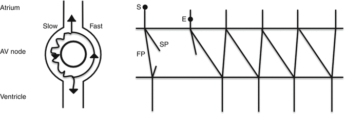
Fig. 19.4
Typical AVNRT (slow-fast) mechanism. FP fast pathway, SP slow pathway, S sinus beat, E extrasystoles
Atypical AVNRT (fast-slow variant) initiates with a ventricular premature complex that is blocked in the fast pathway. The impulse then conducts to the atrium via the slow pathway and returns to the ventricle via the fast pathway. Seldom an atrial premature complex initiates tachycardia (Fig. 19.5).
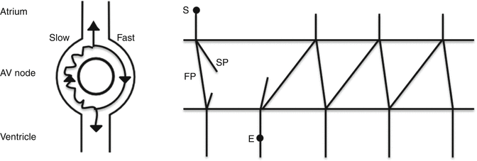
Fig. 19.5
Atypical AVNRT (fast-slow) mechanism. FP fast pathway, SP slow pathway, S sinus beat, E extrasystoles
In AVNRT, neither the atrium nor the ventricle has a critical role in the reentrant circuit [6].
Clinical Presentation and Diagnosis
AVNRT usually has a sudden onset and termination, without a warm-up period. Its symptoms include palpitations, neck pulsations, dizziness, and in some cases chest pain or dyspnea. Tachycardia can last a few minutes or hours, and the termination can be spontaneous or not. Sometimes precipitating factors (drugs, physical and psychological stress, menstruation, anemia, hyperthyroidism, etc.) can be found [5–7].
A 12-lead ECG is fundamental for the diagnosis. ECG, during AVNRT, shows in most cases a regular narrow QRS complex tachycardia; sometimes the QRS complex can be wide with the typical bundle branch block morphology (LBBB or RBBB). Rate of tachycardia is more often between 140 and 250 per minute. The AV relationship is practically in all cases 1:1. Atrial activation is concentric type because the retroconducted stimulus comes from the AV node, so the retroconducted P wave is narrow and negative in inferior leads. In typical AVNRT, the impulse conducts to the atrium via the fast pathway so the atrium and ventricle are activated simultaneously during tachycardia; the retrograde P wave is either hidden within the QRS or embedded in the terminal portion of the QRS, resulting in RP < PR (usually RP is less or equal to 70 ms) with pseudo-r waves in lead V1 and pseudo-s waves in inferior leads. Comparison with the QRS in sinus rhythm can help in identifying pseudo-r and pseudo-s waves. In atypical AVNRT, the conduction to the atrium is through the slow pathway; the atrium is activated late relative to the ventricle, so the retroconducted P wave is away from the previous QRS and it is near the following QRS (RP > PR) [6].
Invasive electrophysiological investigation (EPS) allows to detect the presence of dual AV nodal pathways, to induce and identify AVNRT (typical or atypical), and to perform catheter ablation of tachycardia. Dual pathway can be demonstrated with delivery of a premature atrial complex that results in a “jump” in AH conduction curve (defined as an increase >50 ms in AH interval with a 10 ms increase in atrial extrastimulus prematurity). This “jump” is due to antegrade block in the fast pathway with the conduction through the slow pathway. The electrophysiological characteristic of AVNRT is the concentric retrograde activation of the atrium, with a short ventriculoatrial (VA) interval or with a “A on V” (simultaneous activation of the atrium and ventricle) in typical AVNRT or with a long VA time in atypical (fast-slow) AVNRT [6].
Differential Diagnosis
Surface ECG can make differential diagnosis between AVNRT and the other narrow QRS complex tachycardia during tachycardia, by the induction and termination of tachycardia and by the response to vagal maneuvers [5].
ECG features to assess are shown in Fig. 19.6.
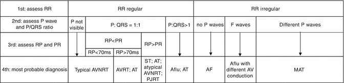
Fig. 19.6
ECG features to assess. AVNRT atrioventricular node reentrant tachycardia, AVRT atrioventricular reentrant tachycardia, AT atrial tachycardia, ST sinus tachycardia, PJRT permanent junctional reciprocating tachycardia, Aflu atrial flutter, AF atrial fibrillation, MAT multifocal atrial tachycardia
The response to vagal maneuvers or to adenosine can help to distinguish between the different types of supraventricular tachycardia. Gradual slowing then reacceleration of rate is typical of sinus tachycardia or focal atrial tachycardia. Sudden termination is typical of AVNRT and AVRT. Persisting tachycardia with transient high grade of AV block is typical of atrial flutter or atrial tachycardia [5].
Treatment
Acute treatment: patients with hemodynamic compromise should be electrically cardioverted without delay. Usually, AVNRT does not cause significant hemodynamic compromise. Vagal maneuvers (Valsalva maneuver, dive reflex, carotid sinus massage) can be used to terminate the tachycardia. These maneuvers increase the vagal tone resulting in slow pathway conduction blockage. If vagal maneuvers are ineffective, medications known to prolong refractoriness of the slow pathway of the AVNRT circuit can be used. These agents include adenosine, calcium channel blocker (verapamil or diltiazem), and beta-blockers (metoprolol, atenolol, propranolol, esmolol). Intravenous administration of these drugs should be performed with continuous ECG monitoring. These agents are very effective in terminating the AVNRT [5, 6].
< div class='tao-gold-member'>
Only gold members can continue reading. Log In or Register to continue
Stay updated, free articles. Join our Telegram channel

Full access? Get Clinical Tree


