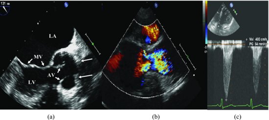
Transesophageal echocardiography (TEE) recording at angle 131° illustrates a diffuse area of supravalvular narrowing (hourglass lesion) with a superimposed membrane. The color Doppler recording shows the blood flow through the narrow part with high velocity (400 cm/sec; gradient is 64 mmHg) from apical 5-chamber view (Figure 50.2, Videoclip 50.1).
Figure 50.2 Transesophageal echocardiography recording at an angle of 131° illustrates a diffuse area of supravalvular narrowing (hourglass lesion) with a superimposed membrane (long arrows) (a). The color Doppler recording shows the blood flow through the narrow part with high velocity (b). The peak velocity of the flow through the narrow part is 400 cm/sec; gradient is 64 mmHg from apical 5-chamber view (c). AV, aortic valve; LA, left atrium; LV, left ventricle; MV, mitral valve.
Stay updated, free articles. Join our Telegram channel

Full access? Get Clinical Tree


