Fig. 17.1
SEPS port placement
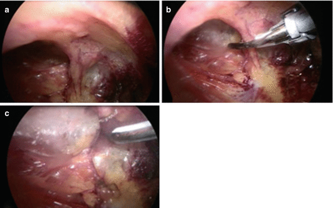
Use of a thigh tourniquet and Esmarch bandage to exsanguinate the limb is reported by some authors [1]. The deep facia is incised under vision and subfacial plane for the port is developed by blunt dissection. Ten-millimeter port is introduced, CO2 is insufflated at 30 mmHg and subfacial space is bluntly created using the scope. Alternatively a balloon insufflation can be used to create this subfacial plane [1]. The second port is now placed 3 cm posterior and a little caudal to the first port. The two ports can be interchanged to facilitate dissection later. All the perforating veins along the medial aspect of the tibia, from the entry of the port till the medial malleolus, are identified, skeletonized, and disconnected. This can be achieved by clipping, diathermy, or use of harmonic scalpel (Fig. 17.2).


Fig. 17.2
(a) Perforator skeletonized. (b) Control with harmonic scalpel. (c) Disconnected perforator
We prefer to use harmonic scalpel due to less trauma, better occlusion of vein, and less incidence of nerve injury. Dissection at the malleolar area is challenging due to lack of space and difficulty in identifying the perforating veins especially the lateral and anterior ankle perforators and the lower most medial calf perforator. Posterior dissection is continued as far beyond midline as possible.
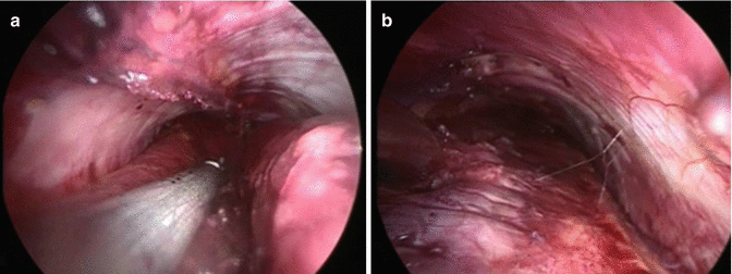
Ports are interchanged for the next part of the dissection. The intermuscular septum under the tibia is identified and opened to gain access to the lateral perforators (Fig. 17.3).


Fig. 17.3
(a) Intermuscular septum divided. (b) Subfascial compartment on completion
These are also disconnected. During this step, the upper two medial calf perforators may be encountered passing within the intermuscular septum and need to be controlled before dividing the septum. A survey is done to ensure completion and hemostasis. Ports are removed and wounds closed. Compression bandages are applied and early ambulation is encouraged. Patient is discharged the next day. This procedure can be combined with HL/S or endovenous ablation and phlebectomy if needed.
Postoperative Complications
There are no major complications reported following SEPS and the procedure is considered extremely safe. There are reports of minor events such as bleeding and hematoma in the subfascial plane, infection in the subfascial plane requiring drainage [15], and muscle herniation. Our experience with the technique is extremely satisfying but the numbers are too small.
Comparison of Open and Endoscopic Perforator Surgeries
The advantages of SEPS over the open procedure are obvious. Delayed wound healing, skin necrosis, and wound infections are common after the open surgery. With SEPS postoperative pain is much less and the patient can be mobilized in the immediate postoperative period. All these would result in a significantly shorter hospital stay for the SEPS group. There are several limitations advanced against SEPS [8]:
Potential risk of injury to posterior tibial artery, vein, and nerve in the distal leg. This is overcome by identifying and confirming the morphology of the perforating veins before dividing them.
Use of electrocautery in the subfascial space can aggravate vascular and nerve injuries. The perforators are clipped and divided rather than controlling them by electrocoagulation. A better option would be the harmonic scalpel.
Expanding the subfascial space in the presence of venous lipodermatosclerosis can be difficult. This can result in missing some of the important perforators. A paratibial fasciotomy can overcome the access problem [1].
The advantages of the endoscopic technique in terms of wound complications are very evident, 0 % for SEPS and 53 % for open surgery. In view of the unacceptably high incidence of wound complications, in a prospective trial, Pierik et al. had to stop their open surgery arm prematurely [8].
Outcome of Perforator Vein Surgery
The outcomes of perforator surgeries, both open and SEPS, have been reported by a number of authors. The mean ulcer recurrence rate following open surgery was 23 %; for SEPS it was 12%. The most obvious and striking difference was in the incidence of wound complications; for open surgery it was 25 %, whereas for SEPS it was less than 7 % [1, 8]. Since the open surgery is now practically obsolete, these data have no relevance.
Clinical Outcome of SEPS
Since SEPS is emerging as the recommended treatment for incompetent perforators, it would be prudent to compare its outcome in the different clinical classes of patients and also in patients with the post-thrombotic syndrome.
Several studies have analyzed the outcome of SEPS in patients with the clinical class of C2 patients. RCTs have concluded that at 1 year in class C2 patients, no additional clinical benefits could be observed when SEPS was combined with HL/S of the GSV [5].
The outcomes in patients of the C3–C6 classes (mostly C5 and 6 classes) are promising. The North American registry reporting on the midterm results of SEPS (NASEPS) documented a cumulative ulcer healing rate of 88 % at 1 year and a cumulative ulcer recurrence rate of 16 % at 1 year, and 28 % at 2 years [16].
Ten Brook and colleagues in a systemic review in 2004 reported ulcer healing rate of 88 % and recurrence of 13 % in a short-term follow-up of SEPS, with or without saphenous ablation [17].
Ulcer healing was considerably delayed in those patients who underwent SEPS alone, in comparison to the group who had SEPS combined with saphenous ablation. The 90-day cumulative ulcer healing rates for the two groups were 49 and 90 %, respectively. Ulcer recurrence at 5 years was also much higher for the SEPS alone group (53 %) in comparison to the other group (19 %) [1].
Stay updated, free articles. Join our Telegram channel

Full access? Get Clinical Tree


