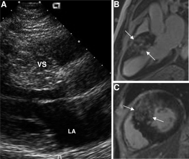
We found a highly inconsistent relation between the granular and reflective ultrasound (“speckling”) pattern frequently observed in the ventricular septum of patients with hypertrophic cardiomyopathy and evidence of myocardial fibrosis by contrast-enhanced cardiovascular magnetic resonance imaging. Therefore, this distinctive echocardiographic appearance of the myocardium does not accurately characterize left ventricular scarring and is most likely explained as an extraneous ultrasound signal pattern. In conclusion, myocardial fibrosis in patients with hypertrophic cardiomyopathy is most reliably identified using contrast-enhanced cardiovascular magnetic resonance imaging.
The tissue characterization of the thickened ventricular septum in hypertrophic cardiomyopathy (HC) has been of interest to students of this disease since the introduction of echocardiography in the early 1970s. Indeed, in the initial 2-dimensional echocardiographic study of HC, ultrasound signals creating a unique granular pattern of myocardial reflectivity with multiple echodensities was noted in the septum of some patients.
Among other possibilities, it has been suggested that this “speckled” appearance might represent an ultrasonic signal marker for the structural properties of myocardium, such as replacement fibrosis, a common finding on autopsy examination of patients with HC. However, image processing studies using integrated backscatter systems to characterize myocardial tissue have not conclusively determined whether the granular and highly reflective (“speckled”) ultrasound appearance of the septum in patients with HC represents areas of scar formation. The introduction of contrast-enhanced cardiovascular magnetic resonance (CMR) imaging with late gadolinium enhancement (LGE) to HC has made it possible to noninvasively identify areas of myocardial replacement fibrosis in vivo and potentially resolve this question in patients with HC.
Patient Images
We studied 38 consecutive patients with HC (aged 44 ± 20 years, 74% men, ventricular septal thickness 23 ± 5 mm), using both echocardiograms and contrast-enhanced CMR studies obtained on the same day. We compared the same imaging planes and divided the patients into 2 subgroups: (1) 20 patients with echocardiograms performed using standard and consistent gain and brightness settings with commercially available instruments, showing a particularly prominent granular (“speckled”) appearance of the basal anterior ventricular septum that were compared to the contrast-enhanced CMR studies; and (2) 18 patients with contrast-enhanced CMR studies showing marked (usually transmural) LGE of the anterior septum that were compared to their echocardiograms.
Of the 20 patients in the first group selected for the “speckled” appearance of the septum on the echocardiogram, LGE was present in the same region of the left ventricular chamber in only 8 (40%; Figure 1 ) and absent in the other 12 ( Figure 2 ). Of the 18 patients in the second group selected for marked LGE of the septum, multiple and prominent echodensities, consistent with “speckling,” were present in the same region of the septum by echocardiography in only 6 (33%) and absent in the other 12. Therefore, in the overall study cohort, the concordance of multiple echodensities with LGE on CMR studies was evident in only 14 (37%) of 38 patients.


