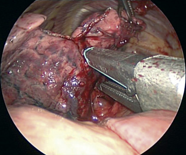
CHAPTER 14 Sleeve Lobectomy
Right Middle Lobe Superior Segment – Video 14
Approach to Video-Assisted Sleeve Lobectomy of the Right Middle Lobe Superior Segment
 Video-Assisted Sleeve Lobectomy of the Right Middle Lobe Superior Segment
Step 1. Right Middle Lobe Vein
♦ The thoracoscope is aimed anteriorly with the 30-degree lens pointed posteriorly toward the hilum.
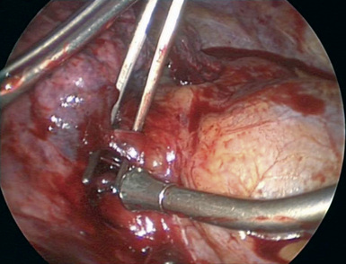
♦ Dissect between the inferior and superior pulmonary veins to define the inferior border of the RML vein and to determine whether there is drainage from the middle lobe to the inferior pulmonary vein (Figure 14-2).
♦ With the vascular endoscopic stapler introduced from the utility incision or the auscultatory incision, transect the RML vein.
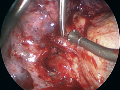
Figure 14-2 Exposure for the right middle lobe vein, superior pulmonary vein, and inferior pulmonary vein.
Step 2. Middle Lobe Artery
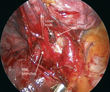
♦ The RML artery parallels the bronchus and is located just superior and slightly medial to the bronchus (Figure 14-4).
Step 3. Minor Fissure
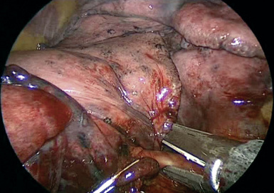
♦ Pull the middle lobe and upper lobe inferiorly to line up the minor fissure with incision 1 or incision 3.
Stay updated, free articles. Join our Telegram channel

Full access? Get Clinical Tree


