Sleep-Disordered Breathing


Sleep-Disordered Breathing


Barbara Phillips


Sleep-disordered breathing is associated with several potentially serious conditions that include:


  • 1. Primary snoring


  • 2. Upper airway resistance syndrome (UARS)


  • 3. Obstructive sleep apnea/obstructive sleep hypopnea syndrome (OSAHS)


  • 4. Central sleep apnea


  • 5. Asthma


  • 6. Chronic obstructive pulmonary disease (COPD).

Each of these conditions has specific clinical presentations with important implications for diagnosis, treatment, morbidity, and follow-up.


PRIMARY SNORING

Primary snoring (or snoring without sleep apnea) is a complex phenomenon that occurs from an interaction between the various upper airway muscles (i.e., tongue, soft palate, and pharynx) and the compliance of the airway walls. Snoring is usually an inspiratory sound, but it can also be noted on expiration. It occurs in all stages of sleep, but most frequently during stages 2 to 4. Vibration of these membranous parts of the airway creates a diffuse involvement of the airway that makes successful treatment of snoring difficult (1).


Clinical Presentation

Patients who present with snoring are commonly referred to an otolaryngologist, usually after complaints from their bed partners about being kept awake by the loud snoring. Unlike subjective complaints of patients with OSAHS, patients with snoring do not suffer from daytime somnolence or sleepiness, insomnia, or sleep disruption (1). However, the patient who snores may also be asymptomatic and unaware of this problem.


Classification

The second edition of the International Classification of Sleep Disorders (ICSD, 780.53-1) defines primary snoring as a “respiratory sound generated in the upper airway during sleep that typically occurs during inspiration or expiration” (2).


Epidemiology

The Wisconsin Sleep Cohort Study reports habitual snoring in about 24% of adult women and 40% of adult men (3). Snoring is the most common symptom noted in breathing disorders such as OSAHS, although not all patients who snore have obstructive sleep apnea (4). Snoring may be a precursor to the development of OSAHS, but it is not in itself a predictor of OSAHS.

Snoring occurs more often in men. Th is male predominance observed in epidemiological studies remains unexplained. The prevalence of snoring appears to increase with age, although some studies have demonstrated a reduction in snoring in patients over 60 years (5,6). Earlier studies on snoring indicate that it may be an independent risk factor for hypertension (5); unfortunately, these studies did not include polysomnography and thus may include patients with frank sleep apnea. It remains unknown whether snoring is a marker for sleep apnea, is a risk factor for cardiovascular and cerebrovascular disease, or results in daytime dysfunction (1). Most likely, snoring is a mild form of sleep-disordered breathing (SDB).


Diagnostic Evaluation

There is no accepted or uniform way to evaluate snoring. When snoring is present together with symptoms of daytime sleepiness and questionable apneas, a diagnostic polysomnogram may be appropriate.


Diagnosis

Snoring is a symptom that must be explored with a review of medical history and performance of a physical examination to determine the predisposition to sleep apnea, the need for a sleep study, and the need for more information by the patient. Snoring is no longer just considered a “noise.” If snoring is causing sleep disturbances including arousals and daytime symptoms like sleepiness, its underlying pathology should be investigated (1).


History

Part of the assessment is to determine if the snoring is present and is causing a sleep disruption for the patient. Table 2.1 outlines helpful hints on collecting a sleep history. Patients seek help for snoring for a variety of reasons. Daytime sleepiness is the most common patient complaint. Commonly, it may be that the bed
partner is bothered by the snoring and is experiencing sleep difficulties themselves. The focus in treatment is based on whether the patient has daytime symptoms and other features associated with SDB. A thorough history should include a review of risk factors associated with primary snoring. Known risk factors are listed in Table 2.2.


TABLE 2.1 Essential Elements of the Sleep History for Sleep Disordered Breathing


  1. Obtain the history in the presence of the bed partner, since the patient may not be aware of their snoring patterns.


  2. Inquire whether the snoring occurs nightly, in certain sleeping positions, or is associated with breathing pauses or gasping.


  3. Inquire about risk factors, including obesity, recent weight gain, use of alcohol, seasonal allergies, nasal congestion, and use of tobacco.


  4. Ask about daytime function. Use a standard questionnaire such as the Epworth Sleepiness Scale (Appendix A) to identify daytime somnolence or the Fatigue Severity Scale to assess fatigue.


  5. Ask about systemic diseases such as hypothyroidism or acromegaly that may increase the likelihood of snoring. Inquire as to any previous surgeries or trauma to the upper airways that may affect airway function and patency.


From Hoffstein, V. Snoring. In: Kryger M, Roth T, Dement WC, eds. Principles and Practice of Sleep Medicine. Philadelphia: Saunders; 2000:813-826.


TABLE 2.2 Risk Factors for Primary Snoring


  • Obesity


  • Alcohol consumption prior to sleep


  • Tobacco use


  • Sedative, tranquilizer, or muscle relaxants


  • Hypothyroidism and other medical conditions


  • Supine sleep position


  • Nasal obstruction and congestion


  • Abnormal upper airway anatomy


Physical Examination

The presence of obesity (BMI >30 kg/m2), increased neck circumference (>16 inches in a female or >17 inches in a male), abnormal airway anatomy (i.e., enlarged tonsils, nasal septal deviation), evidence of cardiovascular status (i.e., pulse rate, blood pressure) should be documented during the physical examination. An examination of the oral pharynx is useful to the treatment of snoring if the airway is small or crowded, or if inflammation is present (1). However, a detailed examination by an otolaryngologist using fiberoptic nasendoscopy may be helpful in snorers presenting with these symptoms. The Mallampati classification can be very helpful in evaluating airway size (Fig. 2.1). Th is classification is a relatively simple grading system, which involves preoperative ability to visualize the tonsillar pillars, soft palate, and base of the uvula. It was designed as a means of predicting the degree of difficulty in laryngeal exposure (7). More recently, it has been shown that a high Mallampati score and nasal obstruction are associated risk factors for obstructive sleep apnea (8).


Differential Diagnosis

The decision to perform further assessments including laboratory polysomnography is based on whether the patient has symptoms of sleepiness or hypertension (Fig. 2.2). A patient who presents with simple snoring is usually a healthy individual with no other symptoms and no anatomic abnormalities. The patient generally presents at the request of his or her family. When no other symptoms are present, this patient would require no further investigations, except for treatment for snoring in the form of weight loss, avoiding sleep in the supine position (positional therapy), or the use of an oral appliance.

A symptomatic patient presents with snoring and other complaints, including:


  • Unrefreshing sleep


  • Excessive daytime sleepiness


  • Poor or reduced performance


  • Headaches


  • Difficulty concentrating or attentional deficits.

Further examination of symptoms related to snoring is conducted in two ways: airway assessment and nocturnal polysomnography. If the symptomatic patient
has an abnormal airway anatomy, he or she may need additional evaluation for possible surgical intervention.


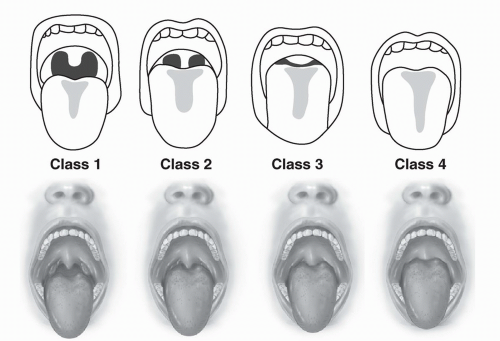
FIGURE 2.1 The Mallampati classification describes tongue size relative to oropharyngeal size. The test is conducted with the patient in the sitting position, the head held in a neutral position, the mouth wide open and relaxed, and the tongue protruding to the maximum. The subsequent classification is assigned based upon the pharyngeal structures that are visible.

Class I = visualization of the soft palate, fauces, uvula, anterior and posterior pillars;

Class II = visualization of the soft palate, fauces, and uvula;

Class III = visualization of the soft palate and the base of the uvula;

Class IV = soft palate is not visible at all.

If the patient phonates, this falsely improves the view. If the patient arches his or her tongue, the uvula is falsely obscured. The test was initially adapted to predict ease of intubation but can be used to predict the potential severity of obstructive sleep apnea (OSA). (See references 7 and 8.) (Modified after Mallampati SR, Gatt SP, Gugino LD, et al. A clinical sign to predict difficult tracheal intubation: a prospective study. Can Anaesth Soc J 1985;32:429-434.)

Nocturnal polysomnography should be considered in the following patients:


  • Asymptomatic snorers contemplating a surgical treatment for snoring


  • Asymptomatic snorers with known vascular disease, including hypertension


  • Asymptomatic snorers, as they may have UARS or OSAHS.

Upper airway resistance syndrome is a term used to describe patients with primary snoring who present with daytime symptoms similar to those noted in OSAHS. UARS describes an increase in airway resistance and a reduction in airflow that does not satisfy the criteria for hypopnea or apnea (9). UARS is present if upper airway resistance is documented within a laboratory investigation and is associated with sleep fragmentation and daytime dysfunction. It should be noted that not every person complaining of snoring has UARS, especially if they present asymptomatically.


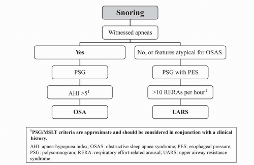
FIGURE 2.2 Algorithm for the evaluation of snoring.


OBSTRUCTIVE SLEEP APNEA-HYPOPNEA SYNDROME

Patients with obstructive sleep apnea-hypopnea syndrome suffer brief episodes of asphyxia during which the oxygen saturation decreases while carbon dioxide levels (CO2) increase (10). After respiration resumes, the patient usually restores normal oxygen and CO2 levels through several recovery breaths or “catch-up breaths.” These episodes, as well as symptoms of snoring, may be witnessed by a family member or bed partner. The patient experiencing obstructive sleep apnea may not be aware that they are apneic or may not remember waking from sleep to resume breathing. Sleep-disordered breathing conditions, such as obstructive apnea and hypopnea, are characterized by complete or partial obstruction of the pharynx during sleep and may result in arousal due to apnea or airflow interruption hypoxemia, or a combination of both. Th is condition can be dangerous and should be investigated and treated promptly.


Clinical Presentation

Patients with OSAHS present with daytime and nocturnal symptoms. Daytime symptoms may include excessive sleepiness, headaches, poor concentration, fatigue, decreased attention, and depression. Nocturnal symptoms include snoring, witnessed apneas, choking episodes, nocturnal dyspnea, restlessness, diaphoresis, nocturia, acid reflux, and drooling. Women with sleep apnea may present differently than their male counterparts; they are more likely to have insomnia, thyroid disease, and depression (11).


Classification

New diagnostic codes and scoring criteria for SDB have recently been published (2,12), including a revised American Academy of Sleep Medicine (AASM) scoring manual.

Classically, apnea is defined as interruption in airflow for a minimum of 10 seconds. Currently, apnea is more specifically defined as a reduction in the amplitude of the thermocouple airflow signal to 90% less of baseline.


The revised AASM scoring manual now describes two definitions for hypopnea.

The “classic” definition of hypopnea, which requires a nasal pressure transducer, is:


a reduction in nasal pressure excursion by >30% of baseline, lasting at least 10 seconds, with a >4% oxyhemoglobin desaturation, with at least 90% of the event meeting the amplitude reduction criterion. An additional definition of hypopnea is a reduction in nasal pressure excursion of at least 50%, lasting at least 10 seconds, with a 3% desaturation or an arousal, with at least 90% of the event meeting the amplitude reduction criterion.

The manual also defines respiratory effort-related arousals (RERA) using nasal pressure (they were originally defined using esophageal pressure) as:


a sequence of breaths lasting at least 10 seconds, characterized by increasing respiratory effort or flattening of the nasal pressure waveform leading to an arousal from sleep when the sequence of breaths does not meet criteria for an apnea or hypopnea.

According to the AASM manual, OSAHS exists when a patient has five or more obstructed breathing events per hour of sleep with the appropriate clinical presentation (12). The expanded definition of hypopnea and the potential for inclusion of RERA in the apnea-hypopnea index (AHI) will inevitably lead to an increased prevalence of sleep apnea and to increased interrater and laboratory variability.

OSAHS is defined as an AHI of at least five apneas plus hypopneas per hour of sleep together with complaints of persistent daytime sleepiness (13). Several medical insurance companies require an AHI of 15 or greater to provide reimbursement for the patient’s treatment using CPAP.

Only gold members can continue reading. Log In or Register to continue

Stay updated, free articles. Join our Telegram channel

Jun 20, 2016 | Posted by in RESPIRATORY | Comments Off on Sleep-Disordered Breathing

Full access? Get Clinical Tree

Get Clinical Tree app for offline access