Sleep and Pregnancy


Sleep and Pregnancy


Christopher Morgan

Hrayr Attarian


The frequency of altered sleep during pregnancy has been reported to range from 13% to 80% in the first trimester and increase to 66% to 97% by the third trimester (1,2). Not all sleep disturbances are due to normal physiologic changes, and a primary sleep disorder should not be overlooked. Although nearly all women admit to poor sleep by the end of pregnancy, only a third believe they have a current sleep “problem” (1), and women who experience sleep problems have a lower perception of general well-being (3). Therefore, a careful sleep history should be taken, as the typical symptoms of a pathologic sleep disturbance may overlap with the normal changes of pregnancy.


SLEEP ARCHITECTURE AND HORMONES DURING PREGNANCY


Normal Sleep Architecture

At the beginning of pregnancy, total sleep time (TST) increases by 30 to 60 minutes, returns to prepregnancy amounts during the second trimester, and is significantly reduced in the third trimester. At one end of the spectrum, women report as few as 3 to 4 hours of TST, particularly if there are other children at home who are not sleeping through the night (4). There is a further dramatic drop off in TST during the first week postpartum, in which subjective reports indicate 1.5 hours less sleep (5). Throughout pregnancy, N1 sleep and wake after sleep onset (WASO) increases, and R sleep either decreases or stays the same. Studies regarding slow-wave sleep (SWS) or N3 sleep are controversial. Most show a decrease throughout pregnancy, but a few, albeit smaller and therefore with a problematic power analysis, have reported a constant or increasing N3 (6,7). Sleep efficiency (SE) decreases in the first trimester, normalizes in the second, and decreases again in the third trimester (8). WASO more than doubles by the third trimester to nearly 10% from prepregnancy levels of <5% (9).

Objective sleep, measured by wrist actigraphy or polysomnogram, has been shown to be on average 30 minutes less than self-reported, as brief awakenings can accumulate up to 45 to 60 minutes of lost sleep (4,10, 11, 12 and 13). A few days before labor, sleep becomes fragmented with decreased TST and increased WASO that become progressively worse until labor (14).

Sleep architecture can remain abnormal for up to 5 months in the postpartum period. In general, TST normalizes, but SE decreases dramatically to 77%, especially in novice mothers (8). In addition, N3 begins to increase, and N1 and N2 begin to decrease back to prepregnancy levels (8,15), more so in lactating women (16). R sleep remains stable for breast-feeding women during the first 2 weeks postpartum, whereas it gradually decreases for nonlactating women (16). R-sleep deprivation can lead to postpartum rapid eye movement (REM) rebound and nightmares (17). Finally, the rare studies on cosleeping and bed sharing show decreased N3 and SE, while R sleep is generally not affected.

Table 19.1 summarizes the key changes in sleep architecture throughout pregnancy and postpartum.


Hormones

The dramatic hormonal changes during pregnancy increase sleep disturbance and affect vigilance directly. High levels of progesterone released from the placenta have a soporific effect, leading to feelings of sleepiness and fatigue early on (18). Progesterone also has a thermogenic effect leading to increased body temperature and an inhibitory effect on smooth muscle leading to urinary frequency (18). As the pregnancy progresses, progesterone continues to rise and estrogen starts to increase. Progesterone contributes to an increase in non-REM sleep, and estrogen can reduce REM sleep. Rising estrogen and progesterone produce airway hyperemia and mucosal edema leading to rhinitis, nasal congestion, and elevated Mallampati scores. These effects increase the risk for snoring and obstructive symptoms in those susceptible.


TABLE 19.1 Sleep Changes Typical During Pregnancy and Postpartum


First Trimester


Second Trimester


Third Trimester


Postpartum


Hormonal changes


High levels of progesterone from placenta


Progesterone levels rises more slowly, estrogen levels begin to rise


Progesterone and estrogen levels peak


No progesterone or estrogen; prolactin fluctuates with lactation


Sleep changes


Fragmented sleep from urinary frequency


Sleep less disrupted


Snoring


More wake time during night


Risk of sleep apnea


Risk of hypertension


Sleep disturbance from leg cramps, heartburn, nasal congestion, urinary frequency


More wake time during night


RLS and PLMS


Disrupted sleep from infant care, breast-feeding, primiparous status


Increased awakenings from breast-feeding


Sleep physiology


+/− Less SWS


TST increased


SE decreased


Light sleep and WASO increased


REM decreased


+/− Less SWS


TST normal


SE normal


Light sleep and WASO increased


REM decreased


+/− Less SWS


TST reduced


SE decreased


Light sleep and WASO increased


REM decreased


More SWS


TST normal


SE decreased


Light sleep normalized


REM sleep deprivation with REM rebound


Physical changes


Increased uterus


Daytime symptoms


Daytime sleepiness and fatigue


Morning or evening nausea


More daytime energy, nasal congestion


Fatigue and sleepiness


Discomfort from irregular contractions, shortness of breath, breast tenderness


Fatigue and sleepiness


Take naps


Postpartum depression


Clinical evaluation


Check serum iron and folate


Assess for SDB


Assess for RLS, SDB


Ask about plans for infant sleeping arrangements


Assess for EDS, cognitive dysfunction, postpartum depression


Ask about infant’s sleep and temperament and parenting activities during the night


Adapted from Wolfson (18) .


Many of these changes are protective both for the fetus and the mother. Elevated levels of progesterone are associated with hyperventilation and enhanced respiratory sensitivity to carbon dioxide, which leads to an augmentation of the respiratory drive and increased responsiveness of upper airway dilator muscles to chemical stimuli. This helps to counteract the normal respiratory changes of pregnancy such as decreased functional residual capacity (FRC), decreased oxygen stores, and secondary shunting and hypoxemia (19,20). In the third trimester, high levels of progesterone and estrogen lead to elevated basal levels of prolactin. Cortisol levels increase twofold in later pregnancy and fourfold during labor; however, circadian rhythms of cortisol, thyroid-stimulating hormone, and prolactin are generally maintained (7). Melatonin also increases in later stages of pregnancy. With elevated melatonin and a lower early morning cortisol peak, a low cortisol-melatonin ratio has been associated with sleeping poorly in the third trimester.

In 35% to 80% of women, the after-delivery rapid decrease in all placental hormones can cause postpartum distress or “blues” that can last up to 2 weeks (21).


SLEEP-DISORDERED BREATHING IN PREGNANCY

In pregnancy, normal changes can predispose women to sleep-disordered breathing (SDB), especially those at higher risk beforehand. The goal of this section is to help clinicians identify the signs and symptoms of SDB in pregnancy and the treatment modalities available to reduce risk to both mother and fetus.


Pathophysiology and Respiratory Changes

Rising estrogen levels, as well as an increase in overall body fluids by up to 7 liters, can produce airway hyperemia and edema of the extremities and mucosa, the latter leading to rhinitis, nasal congestion, elevated Mallampati scores, and increased risk of snoring and airway obstruction. In the last trimester of pregnancy, 42% to 45% of women complain of rhinitis and nasal congestion (22). Both estrogen and another pregnancy hormone, relaxin, cause relaxation of smooth muscles and edema leading to increased collapsibility and decreased caliber of the upper airways (23). Pregnant women have significantly smaller upper airways compared to age-matched postpartum or nonpregnant women (24,25). In general, the decreased caliber of the upper airways has been linked to an increased propensity for SDB. Smaller airways lead to prolonged partial airway obstruction during sleep. The prolonged negative intrathoracic pressures during partial airway obstruction causes release of atrial naturetic peptide (ANP), reducing intravascular volume and cardiac output (26). This, among other factors, can decrease blood flow to the placenta and oxygen delivery to the fetus, exacerbating conditions such as preeclampsia, and increasing the risk for growth restriction. Also, ANP inhibits secretion of antidiuretic hormone, leading to diuresis and increased nocturia and resulting sleep fragmentation.

Progesterone induces an increased respiratory drive and minute ventilation. These changes can lead to a natural respiratory alkalosis with a pH of 7.44 at rest
(27,28). In addition, the growing uterus compresses the diaphragm, resulting in decreased FRC by 20% and decreased expiratory reserve volume. Th is leads to shunting, hypoxemia, and reduced oxygen stores, which could easily contribute to nocturnal respiratory disturbances such as SBD as well as compromised oxygen delivery to the fetus.


FIGURE 19.1 Pathophysiology and consequences of SDB in pregnancy. (Adapted from Edwards and Sullivan [26].)

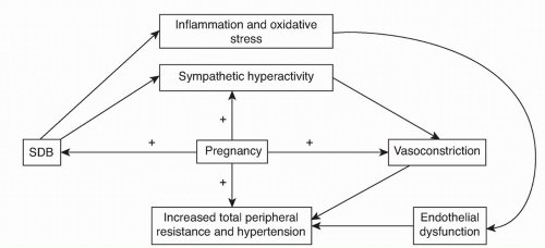
Hemodynamic oscillations associated with apneas are exacerbated during pregnancy (29) and may contribute to maternal complications discussed later. Since sympathetic activity has been shown to be increased in the third trimester of pregnancy (30), SDB may exacerbate this normal physiologic response, which can worsen other comorbitidies such as pregnancy-induced hypertension (PIH) and pre-eclampsia (Fig. 19.1). The increased N1 sleep late in pregnancy may also predispose to increased frequency of central apneas (31).

Some studies on oxygen saturations during normal late pregnancy have found them to be reduced, whereas others found them to remain stable in nonobese women. Saturations, as well as SDB, can be worse in the supine position (19). In addition, the supine position can cause the gravid uterus to compress the vena cava leading to supine hypotensive syndrome, putting the fetus at risk of hypoxemia from uteroplacental insufficiency (32) (Table 19.2).


TABLE 19.2 Physiologic Changes Related to Sleep-disordered Breathing During Pregnancy


Detrimental Changes


Protective Changes


  • Weight gain


  • Elevation of the diaphragm


  • Reduction of functional residual capacity


  • Nasal congestion and rhinitis


  • Hyperventilation


  • Increased stage 1 (non-REM) sleep


  • Sleep fragmentation


  • Avoidance of supine position


  • Increased minute ventilation


  • Increased dilating actions of pharyngeal muscles


  • Reduction of REM sleep


Adapted from Kapsimalis and Kryger (31) .


With the obvious increase in neck circumference and collapsibility of the airway, obesity can significantly worsen obstructive symptoms and increase the risk of SDB in obese mothers (32,33). In nonobese women, normal weight gain during pregnancy has not been proven to confer the same risk (34).

PIH and the more severe forms of pre-eclampsia and eclampsia are associated with increased maternal and fetal morbidity and mortality and have a significant association with SDB (28,29,35, 36, 37 and 38).

Many protective mechanisms also occur. For instance, pregnant women have a preference for sleeping in the lateral position, which may improve cardiac output and oxygenation as well as decrease the frequency of obstructive events (39). An increase in minute ventilation can help to compensate for the increased oxygen demands of pregnancy but can cause symptoms of shortness of breath. As stated before, elevated levels of progesterone increase the responsiveness of upper airway dilator muscles to chemical stimuli, theoretically protecting against airway obstruction. Progesterone may also enhance respiratory drive, increasing negative pressures and causing a tendency for airways to collapse. R sleep is also decreased late in pregnancy, which may help prevent obstructive events since they are more common in this stage (40). Finally, the fetus may be protected by enhanced oxygen delivery by the mother due to a rightward shift in the oxyhemoglobin dissociation curve.


Diagnosis

Screening for SDB should be considered in women with excessive daytime sleepiness (EDS), snoring, apneas, morning headaches, history of intrauterine growth restriction, or in the presence of hypertension or diabetes mellitus (41).

The prevalence of SDB in pregnancy is unknown. Most of the data is from small series and case reports (37,42, 43, 44, 45, 46 and 47) (Table 19.3). Review of these studies suggests that SDB may develop in women with risk factors such as obesity or may worsen the severity of pre-existing SDB, causing maternal and fetal complications discussed later (31,48).

The evolution of SDB over 9 months, peaking during the third trimester, is quite rapid compared to its normal progression over many years. However, the signs and symptoms of SDB in pregnant women are the same for the general population (34). In addition, physical exam findings of obesity, increased neck circumference, and increased Mallampati scores are important factors in the diagnosis of SDB. Women with the above findings should be screened for SDB by overnight polysomnography (PSG) (34).

Women who have PIH and pre-eclampsia risk factors should have meticulous sleep histories taken. Some have suggested that PSG should be performed in those with hypertension alone or previous pregnancies with growth restriction (49), while others feel these are insufficient indications (34). The consensus is that uncomplicated snoring by itself is not an indication for PSG. All of these recommendations are based on uncontrolled or nonrandomized trials and observational studies.


Maternal/Neonatal Complications: Why Do We Need to Treat?


Maternal Complications

The two main maternal complications related to SDB include PIH and gestational diabetes. PIH is a spectrum of disorders ranging from gestational hypertension to pre-eclampsia (proteinuria and edema) to eclampsia (seizures) to HELLP syndrome (hemolysis, elevated liver enzymes, and low platelets). PIH affects about 5% to 10% of pregnancies and is associated with hypertension that does not have the natural nocturnal dip (36). Snoring alone has been linked to increased risk PIH and preeclampsia (28,50, 51, 52 and 53). One possible theory for this association may be the elevated muscle sympathetic activity and nocturnal norepinephrine levels in patients with SDB (54). In pre-eclamptic patients, SDB was associated with increased systemic vascular resistance and suppression of maternal cardiac output (55). Of preeclamptic women, 75% reported snoring and were found to have upper airway narrowing in both the upright and supine positions (25).


TABLE 19.3 Published Reports of SDB During Pregnancy


Author


Year


Number of Cases


Method of Diagnosis


Maternal Complications


Treatment


Fetal Complications


Joel-Coehen and Schoenfeld (133)


1978


3


Clinical


None


None


IUGR


Conti et al. (134)


1988


1


PSG


PIH


None


None


Schoenfeld et al. (135)


1989


8


Clinical


None


None


IUGR


Kowall et al. (136)


1989


1


PSG


PIH, pre-eclampsia


CPAP


N/A


Hastie et al. (137)


1989


1


Clinical


DM


Tracheostomy


None


Charbonneau et al. (138)


1991


1


PSG


DM


CPAP


IUGR


Sherer et al. (139)


1991


1


PSG


DM, PIH (pre-eclampsia)


None


None


Pieters et al. (140)


1995


1


PSG


None


CPAP


None


Lefcourt and Rodis (141)


1996


1


PSG


PIH


None


IUGR


Lewis et al. (142)


1998


1


Clinical


PIH


CPAP


None


Taibah et al. (143)


1998


1


PSG


Hypothyroid


L-thyroxine


N/A


Brain et al. (42)


2001


1 with 2 pregnancies


PSG


PIH, proteinuria


CPAP 2nd pregnancy, 1st trimester


Fetal demise (1st) and IUGR (2nd)


Roush and Bell (46)


2004


1


PSG


PIH


None


IUGR


Guilleminault et al. (43)


2004


12


PSG


PIH


CPAP 1st trimester


None


Louis et al. (47)


2009


57


PSG


PIH (pre-eclampsia)


None


Preterm birth


IUGR, intrauterine growth restriction, IUGR; N/A, not applicable; DM, diabetes mellitus.


Adapted from Kapsimalis and Kryger (31) and Schotland (48) .


Recent studies have shown a strong association of PSG confirmed SDB and gestational hypertension (56). PIH occurs in over 20% of pregnant patients with SDB (37,42, 43, 44, 45, 46 and 47). SDB also has a strong association with gestational diabetes (53), pulmonary hypertension, asthma, and depression (47). Women with pre-eclampsia have an increased prevalence of edema, reduced upper airway caliber, SDB symptoms, and elevated inflammatory markers when compared to healthy pregnant women (25,57).

Despite the above, SDB is a relatively rare cause of these complications in pregnancy (45), and in most cases, causality has not been established. This, however, does not underestimate the importance of screening those with symptoms and risk factors for SDB with PSG, as it is a safe and well-tolerated procedure. Lastly, women with confirmed SDB more commonly have preterm and cesarean deliveries (47), which can cause complications for both the mother and the fetus.


Fetal Complications

Some studies have found no significant difference in mean birth weight, Apgar scores, or complications in newborns (e.g., growth retardation) of snorers compared with nonsnorers (51,54,58), while others demonstrated that habitual snorers were more likely to have infants with lower Apgar scores and growth retardation than nonhabitual snorers (28,52). This discrepancy may be explained by the fact that habitual snoring may be more of a risk than intermittent snoring, as the first set of studies did not differentiate between intermittent snorers and habitual ones.

The frequent apneas, subsequent hypoxemias, repetitive hypertensive peaks, reduction in cardiac output, and increased peripheral vascular resistance that occur in SDB can have detrimental effects on the fetus, including decelerations in fetal heart rate, fetal acidosis, decreased fetal movements from fetal hypoxemia, fetal growth restriction, poor fetal outcomes, and in severe cases, fetal death (28,42,59). It has been hypothesized that reduced fetal and respiratory movements may predispose infants to smaller airways and subsequent development of SDB later in life (26)

Only gold members can continue reading. Log In or Register to continue

Stay updated, free articles. Join our Telegram channel

Jun 20, 2016 | Posted by in RESPIRATORY | Comments Off on Sleep and Pregnancy

Full access? Get Clinical Tree

Get Clinical Tree app for offline access