Sleep and Breathing Assessment


Sleep and Breathing Assessment


Robert Allen and Albert J. Heuer


According to the National Sleep Foundation, approximately 40 million Americans suffer from some type of sleep disorder. The National Heart, Lung, and Blood Institute estimates that of those with sleep disorders, nearly 18 million have some form of sleep apnea. There are more than 70 diagnosed sleep disorders. With 1 in 6 Americans afflicted with sleep-disordered breathing (SDB), respiratory therapists (RTs) must be prepared to identify sleep disorder symptoms in their patients. Critical thinking skills that relate a patient’s known cardiopulmonary history to the presenting complaints and symptoms are key to identifying sleep-related complications or comorbidities.


RTs, sleep technologists, and sleep technicians are important members of the patient care team responsible for conducting a sleep assessments study, or polysomnogram (PSG), and related functions in sleep disorder centers throughout the country. RTs and sleep technicians and technologists working in these centers provide the diagnostic and therapeutic expertise needed to provide primary care physicians, board-certified sleep specialists, and other physician specialists with the information necessary to correctly diagnose and treat sleep disorders.


There are several pathways to entering the sleep profession. Although on-the-job trainees can function without licenses nor credentials in some states, an increasing number of states are requiring credentialing, licensure, or both. At least part of the reason for this is that the Centers for Medicare and Medicaid Services require that only specifically credentialed professionals, including but not limited to RTs, perform sleep studies for Medicare beneficiaries. An individual can be credentialed to perform sleep studies by several boards. The Board of Registered Polysomnographic Technologists grants the credential of Registered Polysomnographic Technologist. The American Board of Sleep Medicine (ABSM) offers credentialing as a Registered Sleep Technologist.


In addition, licensed and credentialed RTs can perform sleep studies, noninvasive positive-pressure titration, and related functions in all 50 states. Many RTs are employed in sleep disorder centers, and their specific background in ventilation, pulmonary anatomy and physiology, pharmacology, and cardiopulmonary diseases makes them valuable assets to the sleep community.


Although ideally providing physical and mental restoration, sleep may be a frustrating experience for many individuals with SDB. Worse yet, sleep disorders can actually exacerbate or compromise preexisting disease conditions during night hours and present life-threatening scenarios. This chapter provides the RT with information on normal sleep staging, the physiologic changes seen during normal sleep, the assessment of SDB, and a brief discussion of selected sleep disorders.


Normal Stages of Sleep


Although dictionary definitions of sleep may suggest a suspension of consciousness and muscular activity or a reduction of one’s awareness of the environment, sleep is actually an active process with continuous stimulation of specific regions of the brain throughout the night.


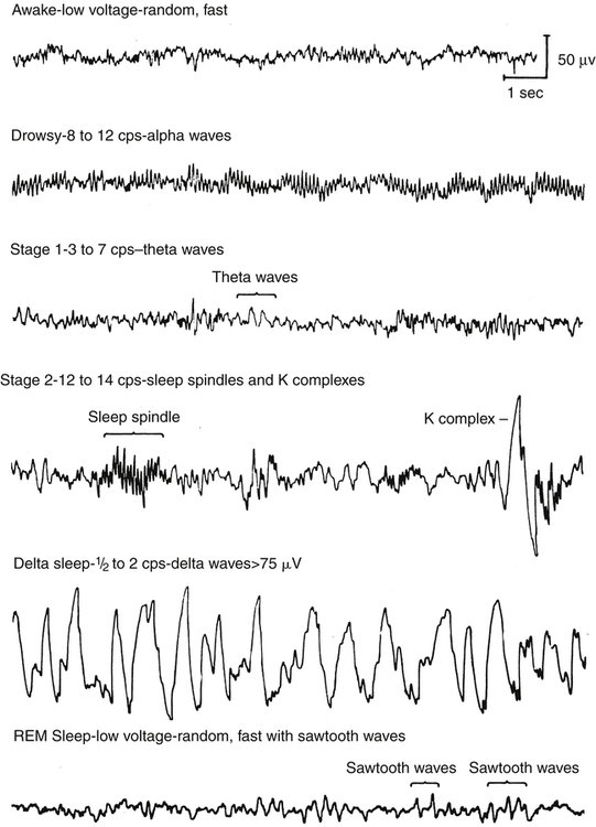
Historically, sleep has been classified in various ways; however, current nomenclature recognizes only two primary states of sleep. For adults and children, the two major states of sleep are non–rapid eye movement (NREM) and rapid eye movement (REM), named for the absence and presence, respectively, of the rapid fluttering or rolling of the eyes and the loss or retention of muscle tone during each phase (Fig. 19-1). There are special states for newborns and infants up to 6 months of age because their brain wave patterns do not yet conform to standard adult classification. They include definitions such as active, quiet, and indeterminate sleep to illustrate the ambiguity of neural activity in the very young. NREM and REM stages cycle back and forth every 60 to 90 minutes for a total of four or five cycles for adults during a normal 8-hour sleeping period. With sleep occupying one third of our life, the quality and quantity of sleep have long-range health and quality-of-life implications.


Non–Rapid Eye Movement Sleep


Sleep normally begins with NREM and progresses to REM sleep. While in NREM, electroencephalogram (EEG) tracings demonstrate diminishing brain activity, suggesting a resting or restorative state. Until recently, NREM was described as four stages of sleep, but a recent reclassification has reduced NREM to three stages. They are differentiated by the amplitude, or wave height, and the frequency, or speed, of the EEG tracings. NREM stages are broken into N1, N2, and N3, the “N” standing for non-REM. With each progressive stage of NREM, the state of sleep is deeper.


As sleep progresses, children and adults normally begin with stage N1. During stage N1, slow rolling eye movements and low-amplitude and mixed-frequency EEG waves are noted. Only 5% to 10% of the sleep period is normally spent in N1. Within 2 to 10 minutes, sleep usually progresses to stage N2. EEG tracings show spikes called K complexes and sleep spindles. In adults, about 40% to 50% of the total sleep period is spent in stage N2.


Stage N3 (formerly stages 3 and 4) is considered the deepest stage of sleep and is about 25% of the sleep period. The EEG demonstrates slow-wave sleep, which is characterized by high-amplitude waves. Delta sleep is hypothesized to be physically restorative sleep. While in N3, sensitivity to external stimuli is diminished, and it is difficult to awaken a person out of this stage of sleep. In addition, the amount of time a person spends in N3 sleep tends to decrease with age, dropping by as much as 50% after puberty. Essential growth hormones are also released during this stage during childhood and adolescence, especially during the first half of the night.


During NREM sleep, the control of core body temperature and regulation of respiration are maintained, the respiratory rate slows, and tidal volume decreases, resulting in an increase in arterial carbon dioxide partial pressure (Paco2). As a result, the inspired minute ventilation is approximately 13% to 15% lower in NREM sleep than during wakefulness. This reduction in minute ventilation for normal human subjects causes an increase in the Paco2 of 2 to 4 mm Hg. Systemic blood pressure may decrease during NREM sleep by 5% to 10% during stages N1 and N2, with a decrease of 8% to 14% during N3 sleep. With age, the percentage of time spent in stages N1 and N2 progressively increases, whereas stage N3 sleep decreases dramatically. Because stages N1 and N2 are lighter stages of sleep, a higher incidence of insomnia is understandably reported with age.


Rapid Eye Movement Sleep


During REM, the brain is active, and dreaming almost always occurs. Throughout the night, REM episodes increase in duration and normally account for about 25% of the total sleep period. EEG tracings demonstrate low-amplitude mixed-frequency waves, similar to those seen in N1 sleep, except that patients in REM have rapid eye movements and a substantial decrease in muscle tone. At their lowest levels during REM, electromyography (EMG) monitoring of muscles of the chin (representative of skeletal muscle tone), demonstrates measurements that are similar to a paralyzed state. This partial paralysis results in a further decrease of the minute ventilation in healthy adults and children, producing associated episodes of hypoxemia and hypercapnia.


The loss of skeletal muscle tone during REM also affects pharyngeal muscles, resulting in increased upper airway resistance as pharyngeal tissues relax and the upper airway lumen is narrowed. Typically, relaxation of the tongue and soft tissues of the oropharynx are the cause of upper airway resistance that may lead to upper airway obstruction. Additionally, heart rate variability is increased, and cardiac arrhythmias are commonly seen during REM.


Characteristically during REM sleep, there is a loss of core body temperature regulation, whereas cerebral blood flow and cerebral temperature increase because of increased brain activity. Systemic blood pressure may become variable and somewhat elevated compared with levels during NREM. Understandably, the physiologic effects of REM may be more profound in patients with preexisting pulmonary or cardiac disease. Normal physiologic changes associated with REM may precipitate increased complications associated with altered ventilation, blood pressure, and heart rate for these particular patients.


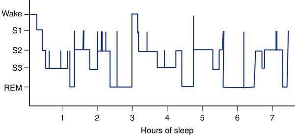
Sleep architecture describes the pattern of various sleep stages that a patient enters throughout the night. Although the approximate percentage of sleep time spent in the three phases of NREM and REM previously described are representative of normal sleep architecture, each person has a distinct pattern particular to his or her own sleep cycle. This pattern can also vary based on the person’s overall tiredness, physical excursion during waking hours, and overall lifestyle.


A histogram of the sleep architecture is part of each sleep study and visually depicts the time spent in each phase of sleep (Fig. 19-2).


In summary, during normal sleep, we begin at stage N1, progress to stage N2, and then reach stage N3 as sleep deepens. During NREM, the brain is in a state of rest, while the body can still move and respond to stimuli. Once REM sleep is initiated, the brain becomes more active, and the body experiences partial skeletal muscle paralysis, with ventilation, blood pressure, and heart rate becoming variable. Although obstructive-type breathing disorders can occur at any stage of sleep, for patients with such a disorder in REM, the soft tissues of the oropharynx relax because of partial paralysis, resulting in upper airway obstruction to airflow. With a loss of airflow and ventilation, the oxygen saturation as measured by pulse oximetry (SpO2) declines, whereas the Paco2 rises. Because the brain is the most sensitive organ to changes in arterial oxygen partial pressure (Pao2) and Paco2, it disrupts sleep at REM onset by “pulling the patient up” out of REM and back into stage N1 or N2 sleep. As the patient then regains muscular control in the NREM state, ventilation is restored, and Pao2, Paco2, and pH values are more normalized; however, sleep has been disrupted to correct this acid-base balance and oxygenation issue. This disruption of sleep to restore oxygenation and acid-base balance is documented as an EEG arousal and usually occurs many times throughout the night.


It is understandable how physically exhausted a patient can become if every time he or she begins the transition into REM, sleep must be interrupted to resume breathing. As a result, such patients report they have not had dreams for years and experience early morning physical exhaustion and headaches as well as excessive daytime sleepiness/somnolence (EDS). Sleep studies in such patients may document 400 to 600 EEG arousals per night and the absence of REM sleep. It should be apparent that when the sleep architecture is disrupted, EDS will result.


A full diagnostic PSG study is required to accurately identify a patient’s sleep architecture. Once documented, RTs, sleep technologists, and sleep technicians can titrate positive airway pressure (PAP) devices to relieve SDB and return the patient to more normalized sleep architecture. The goal in the treatment of all SDB is to minimize arousals and sleep disruptions, helping achieve normalized sleep architecture.


Assessment of Sleep-Disordered Breathing


Results of the physical examination are frequently nonspecific and unremarkable for the patient with SDB. Often, the symptoms of SDB may mimic those of other sleep disorders, leaving it hard to diagnose without proper testing. On inspection, patients most commonly present with obesity and hypertension, although many present with normal body habitus, skin coloring, and respiratory rate and no discernible features of SDB. If a severe and chronic SDB condition does exist, physical examination findings similar to cor pulmonale or congestive heart failure (CHF) may be evident. When conversing with the patient, symptoms of EDS may be present, and if left undisturbed, the patient may begin to quickly doze during an assessment. In some cases, the loss of sleep can produce lethargy or an inability to concentrate on tasks and questions.


By far, the most common problem found in SDB is obstructive sleep apnea (OSA). Sleep apnea is defined as the cessation of airflow for at least 10 seconds during sleep caused by an obstruction in the airway with increased diaphragmatic effort, a change in the EEG, or an EEG arousal, that is at least 3 seconds in length. Sleep apnea can be caused by multiple anatomic and physiologic conditions, and these aspects are discussed later. An EEG is the documented record of brain wave activity collected from electrodes placed on the head and face, using a measuring system referred to as the international 10-20 system, or simply “10-20,” so-named because it uses percentages of 10 and 20 in relation to physical “landmarks” on the human head. The EEG waveforms are used to document brain wave activity and to identify the levels or stages of sleep during a sleep study. An EEG arousal occurs when a patient’s sleep is momentarily disrupted and is documented by a change in the EEG tracings for at least 3 seconds during a sleep study. Many situations may cause an EEG arousal such as a loud noise, room temperature, a dream, body positioning in the bed, or an apnea episode. Patients will not be aware that they have sleep apnea but may comment that they often awaken from sleep with a gasp, snort, or loud snore.


One of the most common findings associated with sleep apnea is snoring. Snoring is the noise produced during inspiration during sleep as a result of soft tissue vibrations in the palate and pillar regions of the oropharynx. Snoring is never a healthy sign, and many snorers may eventually develop significant sleep apnea. Because patients are not aware of their snoring problem, a roommate or family member may be more likely to provide this portion of the medical history. Snoring may be the first sign of a sleep disorder in children and adults. Although families may jest about the snoring of a family member, perpetual snoring is an indicator for a medical examination and possibly a diagnostic sleep study.


Another common symptom associated with sleep apnea, and other SDBs, is EDS, which is difficulty maintaining wakefulness. Unlike sleep apnea, patients with EDS are very conscious of the fact that they have difficulty remaining awake during the day. EDS is a hallmark of sleep disorders. Everyone has experienced the difficulty of staying awake after a night of interrupted or shortened sleep. For individuals with sleep disorders, EDS is likely to be a daily experience that often interferes with the patient’s ability to function. EDS ultimately increases the possibility of workplace or school accidents and has a negative impact on overall productivity.


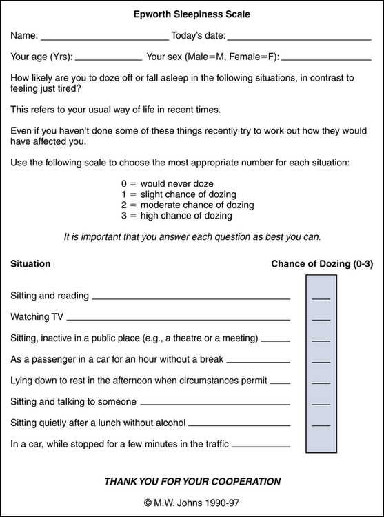
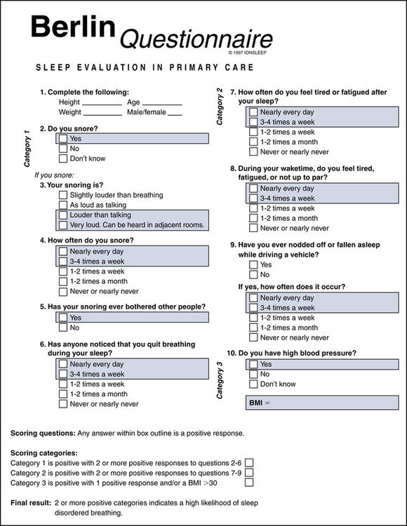
Assessment of snoring and excessive somnolence issues can easily be obtained during the medical history and interview portion of the physical assessment. The inclusion of a few questions regarding snoring and daytime sleepiness will strengthen the assessment process and assist the RT in confirming other health-related issues associated with sleep disorders. Two instruments have been developed that can provide additional valuable information and insight. The Epworth Sleepiness Scale (ESS) is a tool used to assess daytime sleepiness, and the Berlin Questionnaire is a survey instrument used to identify risk factors associated with sleep apnea.


Epworth Sleepiness Scale


The ESS is a simple, eight-item questionnaire that measures daytime sleepiness and is essential for initial screening of sleep disorders. The ESS was developed by Dr. Murray Johns of the Epworth Hospital in Melbourne, Australia in 1991. The survey presents eight situations, and the patient is asked to rate the chances of dozing in each situation, with the value of 0 representing no chance of dozing; 1, a slight chance of dozing; 2, a moderate chance of dozing; and 3, a high chance of dozing. The sleepiness score is totaled, and if the ESS total is 1 to 6, sleep is appropriate and no EDS is noted. If the ESS total is 7 or 8, the score is considered average. An ESS score of 9 or greater indicates that the individual should consult a sleep specialist without delay because there is evidence of EDS (Fig. 19-3). The ESS questionnaire has been shown to have a high degree of reliability and internal consistency as an evaluation instrument.


Berlin Questionnaire


The Berlin Questionnaire is an outcome of the 1996 Conference on Sleep in Primary Care held in Berlin, Germany. More than 120 American and German pulmonary and primary care physicians developed the questionnaire from the literature to identify behaviors consistent with the presence of SDB. The focus of the instrument is deliberately limited to risk factors associated with sleep apnea. The 10-item survey is divided into three categories, with category 1 addressing snoring, category 2 identifying EDS, and category 3 assessing current blood pressure and body mass index (BMI) (Fig. 19-4). To be classified as at high risk for sleep apnea, an individual must qualify for at least two categories. The Berlin Questionnaire has a high degree of sensitivity and specificity for predicting sleep apnea in individuals.


The usefulness of the ESS and the Berlin Questionnaire is critical to the assessment of possible sleep disorder complications in the cardiopulmonary patient. Inclusion of one or both surveys during the patient assessment will provide the RT with information crucial to identifying patient symptoms. A description of signs and symptoms associated with selected SDBs is provided later in the chapter. Before focusing on specifics of SDB, the normal stages of sleep should be examined to better understand the health impact of disrupted sleep.


Jun 18, 2016 | Posted by in RESPIRATORY | Comments Off on Sleep and Breathing Assessment

Full access? Get Clinical Tree

Get Clinical Tree app for offline access