ECG characteristics
The sinus node initiates the electrical impulse that activates atrial and then ventricular myocardium during each normal heartbeat. Sinus node activity itself does not register on the electrocardiogram (ECG).
P wave
Atrial activity, the P wave, is usually apparent in most ECG leads (Figure 1.1). However, occasionally the P wave in some leads is not visible or is of low amplitude, and it may be necessary to inspect all leads of the ECG to establish that there is sinus rhythm (Figure 1.2).
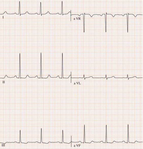
The sinus node lies at the junction of the superior vena cava and right atrium. Atrial activation therefore spreads from the sinus node in an inferior direction (i.e. towards the feet) to the atrioventricular (AV) junction. The P wave, therefore, is upright in those leads that are directed to the inferior surface of the heart (i.e. II, III and aVF), and is inverted in aVR, which faces the superior heart surface (Figure 1.1). If a P wave does not have these characteristics then, even though a P wave precedes each ventricular complex, the sinus node has not activated the atria and the rhythm is abnormal (Figure 1.3).
PR interval
The AV node is the only electrical connection between atria and ventricles: the mitral-tricuspid valve ring that separates the atria from the ventricles is fibrous and cannot conduct electrical impulses. The AV node conducts relatively slowly, thereby delaying conduction of the atrial impulse to the ventricles. Conduction through the AV node does not register on the ECG. The PR interval, which is measured from the onset of the P wave to the onset of the ventricular complex, indicates the time taken for an atrial impulse to reach the ventricles. The normal PR interval ranges from 0.12 to 0.21 s. It should shorten during sinus tachycardia.