Silicosis and Coal Worker’s Pneumoconiosis

Chapter 51 Silicosis and Coal Worker’s Pneumoconiosis


Pneumoconiosis has been defined as the non-neoplastic reaction of the lung to inhaled mineral or organic dust. Prolonged inhalation of coal mine dust may result in the development of pneumoconiosis, silicosis, and industrial chronic bronchitis and emphysema, either singly or in various combinations. Coal worker’s pneumoconiosis (CWP) is the term generally applied to interstitial disease of the lung resulting from chronic exposure to coal dust, its inhalation and deposition, and the tissue reaction of the host to its presence, whereas silicosis refers to lung disease due to inhalation of dust containing silica. Pneumoconioses differ in a number of ways from acute allergic and toxic interstitial diseases associated with exposure to organic dusts, principally because of their long latency periods (usually 10 to 20 years or more) between exposure onset and disease recognition.


Sources of Exposure


Coal is not a mineral of fixed composition. It is graded by rank, reflecting its carbon content and thus combustibility: Anthracite is the highest-ranked coal, with a carbon content of approximately 98%. Lower-ranked coals, bituminous and subbituminous, have carbon contents of approximately 90% to 95% carbon. The rank of coal has an influence on the risk of disease: Higher-rank coals entail higher risk than lower-rank coals. However, exposure to coal dust with a quartz concentration greater than 15% is associated with a high risk for development of a rapidly progressive form of pneumoconiosis that has the characteristics of silicosis. In open mines, dust levels rarely approach those of underground mines.


The most common form of crystalline silica is quartz. Quartz is almost pure silicone dioxide but often contains traces of other elements. Other crystalline forms of silica are cristobalite and tridymite. The importance of silica as a health hazard is due to its ubiquity (Table 51-1). Diatomite is a siliceous sedimentary rock used for filtration; for heat and sound insulation; as an adsorbent and filtering agent; as a filler material in plastics, paper, and insecticides; and in the manufacture of floor coverings.


Table 51-1 Major Industries Associated With Silica Exposure


Occupation Exposure
Sand blaster Ship building, oil rig maintenance, preparing steel for painting
Miner Surface coal mining, roof bolting, shot firing, drilling, tunneling
Miller Silica flour
Glass maker Polishing with sand and enamel work
Potter cleaner Crushing flint and fettling, foundry work, mold making and vitreous enameling, manufacture of cultured quartz crystal
Quarry and stone worker Cutting of slate, sandstone, and granite
Abrasive worker Inhalation of fine particles during grinding

It seems that development and progression of silicosis depend on the total amount of quartz to which workers are exposed, the time over which that exposure occurs, and the presence of other minerals that may interfere with the toxicity of the quartz.


Epidemiology


CWP was first recognized in Scottish miners in 1830. In recent decades, the incidence of CWP has been declining in industrial countries thanks to improved dust controls, although increased mechanization in the mid-1960s led to a temporary increase in dust levels. In parallel, in a report from the United Kingdom, for the period 1950 to 1980, the annual rate of CWP recognition for state compensation in current and retired miners decreased from approximately 7% to 1% to 2%. The overall prevalence of CWP, which reflects more distant exposure and earlier incidence, declined from approximately 13% to 5%, but regional differences in reported rates were substantial. Similar regional differences and similar declines have been noted in the United States and other countries.


Since the 2000s, however, new sources of silicosis have emerged, especially in developing countries—for example, in Turkey, where the denim industry has been responsible for more than 75 cases thus far. Denim sandblasting is used to give jeans a more “worn-out” appearance and requires highly pressurized sand projection, often performed by young persons without any respiratory protection. In a recent epidemiologic study, among 145 workers recruited from the outpatient clinic at Atatürk University, 77 (53%) presented with radiologic evidence of silicosis.


Other, more anecdotal sources of silica exposure have been described, such as heat-dried mud inhalation in workers engaged in the manufacture of tatami mats in China, and handling quartz-containing fillers by dental supply factory workers in the United States. All of these “new forms” of silicosis underscore the fact that it will always remain a concern for respiratory clinicians worldwide, despite the decline of the mining industry in Western countries.


Pathophysiology


Three groups of factors are known to influence the character and severity of lung tissue reaction to mineral dusts. The first category is the intensity and duration of exposure, followed by individual susceptibility, which explains why, among a group of workers exposed to the same dust, only a fraction will develop pneumoconiosis. Finally, the nature and properties of the dust are to be considered. For each mineral, geometric and aerodynamic properties, chemistry, and surface properties vary. Particles that can cause pneumoconiosis are aerodynamically and geometrically small enough to reach the respiratory bronchioles and be deposited there—this generally means spherical particles of 0.5 to 5 µm in diameter.


The pathogenesis of pneumoconiosis is similar to that of all interstitial lung diseases. The condition begins as a chronic inflammatory state (alveolitis) in which inflammatory cells are activated, with consequent damage to the pulmonary architecture. Inorganic particles are phagocytosed by alveolar macrophages, causing their activation and the release of inflammatory mediators such as cytokines and arachidonic acid metabolites. These mediators, in turn, induce the recruitment of other inflammatory cells within the alveolar wall and on the alveolar epithelial surface. Toxic oxygen derivatives and proteolytic enzymes are released by the inflammatory cells, which cause cellular damage and disruption of the extracellular matrix.


The inflammatory phase is followed by a reparative phase, in which growth factors stimulate the recruitment and proliferation of mesenchymal cells and regulate neovascularization and reepithelialization of injured tissues. During this phase, abnormal or possibly uncontrolled reparative mechanisms may result in the development of fibrosis. Fibrogenic particles activate proinflammatory cytokine production within the respiratory tract. Tumor necrosis factor (TNF)-α seems to play a key role in the recruitment of inflammatory cells induced by toxic dusts (Figure 51-1). In addition, neutrophils recruited in the area of inflammation may contribute to the alveolitis, and respiratory and endothelial cells may play a further role by releasing various chemokines such as interleukin (IL)-8. Finally, growth factors such as platelet-derived growth factor, insulin-like growth factor, fibroblast growth factor, and transforming growth factor-β are involved in the pathogenesis of lung fibrosis and in the proliferative response of type II epithelial cells, which occurs in progressive massive fibrosis (PMF).


Histopathologic Changes


CWP lesions are focal. Simple CWP is associated with macular and nodular lesions, whereas complicated CWP is associated with PMF and lesions of rheumatoid pneumoconiosis (Caplan syndrome, discussed later on).


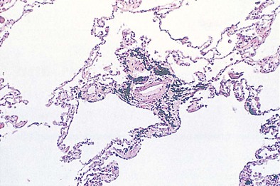
The pleural surfaces of a coal worker’s lung show an irregular pattern of bluish-black pigmentation that corresponds to the junction sites of septal-lymphatic vessels and the pleura. Peribronchial, hilar, and paratracheal lymph nodes are enlarged, black, and firm. The initial lesions in the lung are the coal dust macules, which correspond macroscopically to focal areas of black pigmentation. On microscopic examination, the macule is seen to be composed of coal dust–laden macrophages within the walls of the respiratory bronchioles and adjacent alveoli (Figure 51-2). Focal emphysema around the coal dust macule is common and is considered an integral part of the lesion of simple CWP.


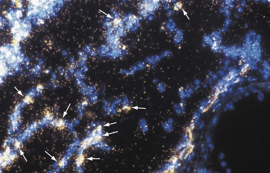
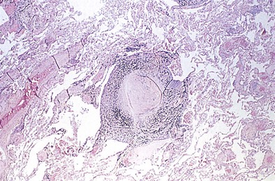
The histopathologic hallmark of simple CWP is the nodule. The nodules are rounded lesions with collagenous centers. On microscopic examination, the nodule can be divided into three zones: a central zone composed of whorls of dense, hyalinized fibrous tissue; a middle zone made up of concentrically arranged collagen fibers (onion-skinning); and a peripheral zone of more randomly oriented collagen fibers mixed with dust-laden macrophages and lymphoid cells (Figure 51-3). “Old” inactive nodules often are relatively acellular. Particles of silica may be demonstrated in the nodules as birefringent particles under polarized light. Nodules represent a form of mixed-dust fibrosis (i.e., coal dust plus silica exposure), usually are found in association with macules, and in some instances may develop from preexisting macules. They are not confined to the respiratory bronchioles but also are seen in the subpleural and peribronchial connective tissues. Nodules tend to cluster and eventually coalesce to produce PMF. Degenerative changes commonly are observed in the nodular lesions, including calcification, cholesterol clefts, and cavitation. In severe silicosis, structural alterations of the pulmonary vasculature may result from the accumulation of dust in the adventitia of large vessels, and involvement of the smaller blood vessels by silicotic nodules also may be seen.


Progressive massive fibrosis

Stay updated, free articles. Join our Telegram channel

Jun 12, 2016 | Posted by in RESPIRATORY | Comments Off on Silicosis and Coal Worker’s Pneumoconiosis

Full access? Get Clinical Tree

Get Clinical Tree app for offline access