Reflux of contrast medium into the inferior vena cava (IVC) is often detected on computerized tomographic pulmonary angiogram. The potential clinical implications and associated diagnoses of this finding have not been established. We investigated the prevalence and significance of reflux of contrast medium into the IVC in a large cohort of patients evaluated for possible pulmonary embolism (PE) by computerized tomographic pulmonary angiography. We retrospectively reviewed 1,065 consecutive computerized tomographic pulmonary angiographic examinations performed from January 1, 2007 through January 7, 2008 for the presence of reflux. Degree of reflux into the IVC and hepatic veins was graded from 1 (none) to 6 (severe). Patients’ charts were reviewed for diagnoses during the index hospitalization and for background diseases. These clinical data were correlated with the reflux grade. The final study included 967 computerized tomographic pulmonary angiographic scans of 367 men and 600 women (mean age 62 ± 20 years, range 17 to 103). Almost 1/2 (480, 49.6%) had grade 1, 310 (32.1%) had grades 2 to 3, and 177 (18.3%) had grades 4 to 6. Multivariate logistic regression found that pulmonary hypertension, history of congestive heart failure, chronic atrial fibrillation, and acute PE were associated with extensive reflux (grades 4 to 6) with odds ratios (95% confidence intervals) of 5.4 (3.0 to 9.9, p <0.001), 3.7 (2.3 to 6.1, p <0.001), 2.3 (1.0 to 5.3, p = 0.044), and 1.8 (1.2 to 2.9, p = 0.011), respectively. Interobserver agreement between the 2 readers for reflux grading was good (kappa = 0.77). In conclusion, extensive reflux of contrast medium into the IVC detected on computerized tomographic pulmonary angiogram may serve as a pathophysiologic marker of right heart dysfunction, specifically pulmonary hypertension, congestive heart failure, chronic atrial fibrillation, or PE.
The number of computerized tomographic pulmonary angiographic (CTPA) examinations performed for evaluating patients presenting with acute dyspnea has been growing exponentially during recent years, with a relatively low prevalence (sometimes even <10%) of findings consistent with the suspected diagnosis of acute pulmonary embolism (PE). Reflux of contrast medium from the right atrium to the inferior vena cava (IVC) and hepatic veins during the first pass of the injected bolus of contrast medium is often present on CT pulmonary angiogram. Only a few preliminary small series have described this finding in association with right heart failure. The aim of the present study was to investigate the prevalence, severity, associated diagnoses, and eventual prognostic significance of reflux of contrast medium into the IVC in a large cohort of patients who underwent CT pulmonary angiography. We considered that these imaging findings might serve as a pathophysiologic marker of right heart dysfunction, thus tapping information obtained by the radiologist to help explain a patient’s clinical presentation.
Methods
The institutional review board approved this retrospective study and waived informed consent. We evaluated 1,065 consecutive CTPA studies performed in 1,027 patients from January 1, 2007 through January 7, 2008. In the event of multiple scans, results of the first scan were entered into the analysis. CT pulmonary angiography is performed at our institution within 12 hours of a clinical suspicion of acute PE, whereas initiation of treatment before the patient undergoes CT pulmonary angiography depends on the degree of clinical suspicion and a patient’s status. In addition to gender and age, patients’ charts were reviewed for the reason for referral to CTPA scanning and final diagnoses during the index hospitalization. Data on background and co-morbid conditions were based on previous medical records, patients’ self-reports, and use of relevant medications. Information on mortality was collected from the database of the Ministry of Internal Affairs of Israel.
All study patients were scanned by a multidetector CT scanner (Mx8000 IDT or Brilliance, Philips Medical Systems, Cleveland, Ohio) with 16- or 64-detector rows. Reconstructed slice thickness was 1.0 to 2.0 mm with an increment of 0.5 to 1 mm. Scans were acquired according to our routine nonelectrocardiographic-gated PE protocol with injections of contrast medium consisting of iodinated contrast material 80 to 100 ml at a concentration of iodine 300 mg/ml (Ultravist, Schering, Berlin, Germany) and at rates of 3 to 4 ml/s. An automated bolus tracking technique was used with a region of interest placed within the main pulmonary artery to optimize visualization of the pulmonary arteries. Five seconds after reaching a threshold of 100 HU at the region of interest, scanning began covering the chest from the lung bases to the thoracic inlet. All scans were obtained in a caudal–cranial direction at end of inspiration during a single breath-hold.
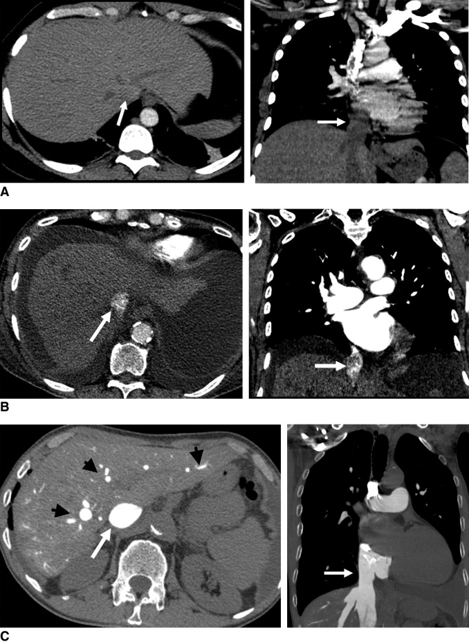
CT scans were reviewed by 2 radiologists (G.A., D.C.) who were unaware of a patient’s clinical history, results of other imaging techniques, and outcome. Severity of reflux of contrast medium into the IVC and hepatic veins was graded from axial images on a scale of 1 to 6 as described by Groves et al (1 = no reflux into the IVC, 2 = trace of reflux only in the IVC, 3 = reflux into the IVC but not the hepatic veins, 4 = reflux into the IVC and opacifying the proximal hepatic veins, 5 = reflux into the IVC and opacifying the midpart of the hepatic veins, 6 = reflux into the IVC and opacifying distal hepatic veins; Figure 1 ). These 6 grades of reflux were decreased to 3 groups for statistical analyses: no reflux (grade 1), mild reflux (grades 2 to 3), and extensive reflux (grades 4 to 6). Grading of reflux of contrast medium into the IVC was performed separately by the 2 radiologists on the first 500 consecutive CT pulmonary angiograms to assess interobserver variation.

Data were summarized as mean ± SD for continuous variables and as number of subjects for categorical variables. These clinical data and mortality data were correlated with severity of reflux. Kappa was calculated to measure agreement between the 2 independent observers for grading extent of reflux. Comparison of frequencies among groups of reflux grades was by chi-square statistics. Univariate and multivariate logistic regression models were used to calculate the odds ratio for having extensive reflux in each diagnosis. We used Cox proportional hazard models to evaluate the hazard ratio of severe reflux for the outcome of mortality. All these analyses were considered statistically significant at a p value <0.05 (2-tailed). SPSS (SPSS, Inc., Chicago, Illinois) was used to perform all statistical evaluations.
Results
Reflux could not be measured owing to technical reasons on 63 of 1,065 scans (5.9%): the hepatic venous confluence was not included in the scan in 33 patients, there was poor enhancement of the right heart and pulmonary vessels in 22 patients, and injection was performed from the femoral vein in 8 patients. Thirty-four patients had multiple CTPA scans, 26 patients had 2 scans, and 4 patients had 3 scans. Thus, the final study group included 967 CTPA scans for 367 men (38%) and 600 women (62%) whose mean age was 62 ± 20 years (range 17 to 103).
The 2 most common reasons for referral were symptoms of dyspnea and/or low oxygen saturation (763 CT pulmonary angiograms, 76.1%) and chest pain (116 CT pulmonary angiograms, 11.6%). PE was diagnosed in 17% of patients, whereas infection and oncologic diseases were the most frequent acute final diagnoses. Baseline characteristics of the study cohort including final diagnoses during the index hospitalization, background, and co-morbid conditions are presented in Table 1 .
| Total | 967 (100%) |
| Women | 600 (62%) |
| Hypertension | 404 (41.8%) |
| Infection | 331 (34.2%) |
| Ever smoker | 284 (29.4%) |
| Dyslipidemia | 231 (23.9%) |
| Diabetes mellitus | 186 (19.2%) |
| Oncology | 184 (19.0%) |
| Ischemic heart disease (acute/chronic) | 183 (18.9%) |
| Acute pulmonary embolism | 167 (17.3%) |
| Chronic obstructive respiratory disease | 164 (17.0%) |
| History of congestive heart failure | 119 (12.3%) |
| Cerebrovascular event | 86 (8.9%) |
| Obesity | 84 (8.7%) |
| Congestive heart failure exacerbation | 79 (8.2%) |
| Previous pulmonary embolism | 53 (5.5%) |
| Chronic atrial fibrillation | 44 (4.6%) |
| Acute respiratory failure | 31 (3.2%) |
| Inflammation | 30 (3.1%) |
| Other | 514 (53.2%) |
There were 480 patients (49.6%) with no evidence of reflux (grade 1), 310 (32.1%) with mild reflux (grade 2 to 3), and 177 (18.3%) with extensive reflux into the IVC and hepatic veins (grade 4 to 6). Tables 2 to 4 present the number and percentage of subjects with each grade of reflux and the age-adjusted odds ratio and 95% confidence interval for having extensive reflux for each diagnosis. Advanced age, most cardiac diagnoses, and obesity were associated with an increased prevalence and odds for having extensive reflux. In contrast, none of the pulmonary diagnoses were associated with extensive reflux, whereas an oncologic diagnosis was associated with a low prevalence of reflux.
| Variable | Reflux | Age-Adjusted OR for Extensive Reflux | ||||
|---|---|---|---|---|---|---|
| None (grade 1) | Mild (grades 2–3) | Extensive (grades 4–6) | p Value | OR (95% CI) | p Value | |
| Age (years), mean ± SD | 59 ± 19 | 64 ± 20 | 67 ± 22 | <0.001 | 1.19 ⁎ (1.09–1.30) | <0.001 |
| Women | 286 (59.6%) | 198 (63.9%) | 116 (65.5%) | 0.105 | 1.20 ⁎ (0.86–1.69) | 0.290 |
| Acute pulmonary embolism | 85 (17.7%) | 46 (14.8%) | 36 (20.3%) | 0.125 | 1.18 (0.78–1.79) | 0.426 |
| Previous pulmonary embolism | 21 (5.0%) | 21 (7.6%) | 11 (6.9%) | 0.153 | 1.24 (0.62–2.49) | 0.541 |
| Acute or history of pulmonary embolism | 98 (20.4%) | 56 (18.1%) | 41 (23.2%) | 0.181 | 1.17 (0.79–1.74) | 0.439 |
| Variable | Reflux | Age-Adjusted OR for Extensive Reflux | ||||
|---|---|---|---|---|---|---|
| None (grade 1) | Mild (grades 2–3) | Extensive (grades 4–6) | p Value | OR (95% CI) | p Value | |
| Congestive heart failure exacerbation | 17 (3.5%) | 23 (7.4%) | 39 (22.0%) | <0.001 | 4.48 (2.74–7.33) | <0.001 |
| History of congestive heart failure | 25 (6.1%) | 33 (12.4%) | 61 (38.9%) | <0.001 | 6.00 (3.87–9.30) | <0.001 |
| Congestive heart failure (acute/chronic) | 34 (7.1%) | 40 (12.9%) | 68 (38.4%) | <0.001 | 5.53 (3.65–8.37) | <0.001 |
| Ischemic heart disease (acute/chronic) | 70 (14.6%) | 70 (22.6%) | 43 (24.3%) | 0.001 | 1.20 (0.80–1.80) | 0.381 |
| Chronic atrial fibrillation | 6 (1.5%) | 10 (3.7%) | 28 (17.6%) | <0.001 | 7.43 (3.87–14.25) | <0.001 |
| Aortic stenosis | 2 (0.5%) | 8 (3.0%) | 3 (1.9%) | 0.006 | 1.01 (0.27–3.75) | 0.985 |
| Aortic regurgitation | 4 (1.0%) | 4 (1.5%) | 7 (4.5%) | 0.004 | 3.26 (1.15–9.19) | 0.026 |
| Mitral stenosis | 2 (0.5%) | 0 (0%) | 6 (3.8%) | <0.001 | 12.89 (2.55–65.03) | 0.002 |
| Mitral regurgitation | 10 (2.4%) | 10 (3.7%) | 15 (9.5%) | <0.001 | 2.93 (1.45–5.92) | 0.003 |
| Tricuspid regurgitation | 5 (1.2%) | 7 (2.6%) | 18 (11.4%) | <0.001 | 6.38 (2.98–13.68) | <0.001 |
| Pulmonary hypertension | 8 (2.0%) | 18 (6.8%) | 45 (28.7%) | <0.001 | 8.99 (5.30–15.26) | <0.001 |
Stay updated, free articles. Join our Telegram channel

Full access? Get Clinical Tree


