5 Shunting Lesions
Atrial Septal Defects
Atrial deptal defects represent defects in septation of the atria. They are common, representing as much as 10% of cases of congenital heart disease. Anatomic types of defects include (Fig. 5-1):
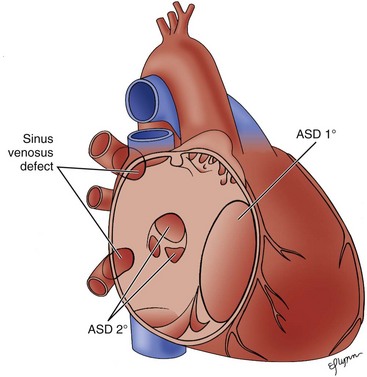
Figure 5-1 Diagram of the atrial septum showing several types of atrial septal defects.
(Adapted from Fyler DC [ed], Nadas’ Pediatric Cardiology. Philadadelphia: Hanley & Belfus, 1992.)
Patent Foramen Ovale
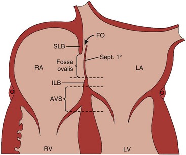
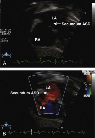
• An interatrial communication between the superior limb of the septum secundum on the right side and the septum primum on the left atrial side (Fig. 5-2).
• Functionally closes after birth but may be present in as many as 25% of normal hearts during pathologic evaluation.
Ostium Primum Atrial Septal Defect
Sinus Venosus Atrial Septal Defects: Two Types
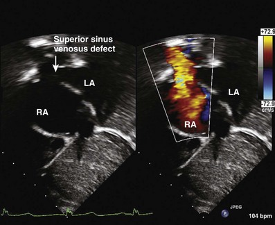
• Superior sinus venosus defects: located in the most superior and posterior region of the atrial septum in close proximity to the right-sided pulmonary veins (PVs) and the entrance of the superior vena cava (SVC) into the right atrium (RA) (Fig. 5-5).
• Inferior sinus venosus defects: located posterior and inferior to the fossa ovalis and adjacent to the inferior vena cava.
The Echocardiography (Echo) Exam: Step-by-Step Approach
Step 1: Evaluate the Location of the Atrial Septal Defect
Key Points
• Dropout can occur in the region of the fossa ovalis, giving the impression that an ASD exists in a normal heart or making an existing defect appear larger when imaged from an apical view.
• Evaluate AVV regurgitation, especially in the presence of a primum ASD or a cleft in the anterior leaflet of the mitral valve.
Step 2: Evaluate the Atrial Septal Defect Dimensions and Position
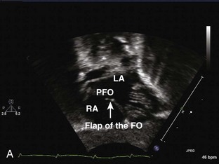
• A PFO is a defect in the septum primum less than 3 mm in size without associated right atrial or right ventricular dilation and typically with a flap of tissue partially occluding the communication.
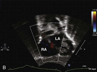
Step 4: Determine the Size of the Shunt
• The left-to-right shunt across an ASD is often minimal during infancy as the RV is initially less compliant.
Post-Device Atrial Septum Defect Closure Evaluation: Special Considerations
Ventricular Septal Defects
A ventricular septal defect is a communication within the interventricular septum that seperates the left ventricle (LV) and RV, allowing for shunting for blood between the ventricles. VSDs represent 20% of congenital heart disease. VSDs can be classified as follows (Fig. 5-6):
Stay updated, free articles. Join our Telegram channel

Full access? Get Clinical Tree


