Shortness of Breath and Dyspnea on Exertion
A 33-year-old man from India complains of shortness of breath and dyspnea on exertion (DOE) walking one to two blocks.
On an exercise test, he exercised for 8 minutes 41 seconds, reaching a heart rate of 181 beats per minute. His pulmonary artery pressure increased from 48 mm Hg at rest to 89 mm Hg after exercise.
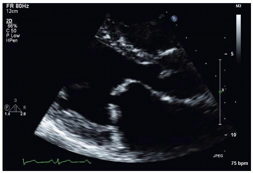 Figure 12-1. |
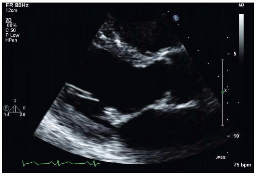 Figure 12-2. |
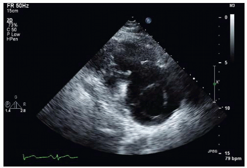 Figure 12-3. |
A. Rheumatic mitral valve disease
B. Rheumatic valvular disease of the aortic valve
C. Rheumatic valvular disease of mitral, aortic, and tricuspid valves
D. Rheumatic mitral and aortic valve disease
E. Degenerative valvular disease
View Answer
ANSWER 1: C. All three valves demonstrate thickening of the leaflet tips with diastolic doming of the mitral and tricuspid valves and systolic doming of the aortic valve. Typically, the mitral valve is most commonly affected followed by the aortic valve, and when the aortic valve is affected, the mitral valve is almost invariably affected. It is much more uncommon to have the tricuspid valve involved.
 Figure 12-4. |
 Figure 12-5. |
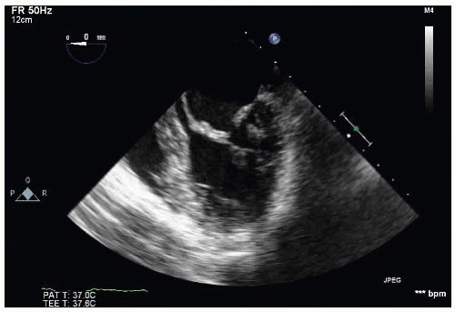 Figure 12-6. |
A. No significant mitral stenosis (MS); significant aortic regurgitation (AR)
B. Significant MS and AR
C. No significant AR; significant MS
D. No significant valvular disease
View Answer
ANSWER 2: B. Figure 12-4 demonstrates a diastolic frame in the parasternal long-axis view, showing severe aortic valve regurgitation: AR vena contracta is greater than 0.6 cm and takes up almost the whole outflow tract. A high velocity jet is seen within the left ventricle, suggesting MS. The significance of the MS is shown in Figure 12-5 using continuous wave across the mitral valve in the apical four-chamber view. The mean diastolic pressure gradient of 11 mm Hg is consistent with severe MS.
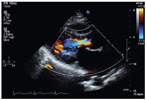 Figure 12-4. |
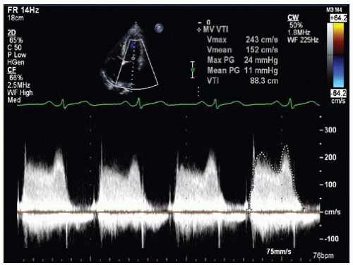 Figure 12-5. |
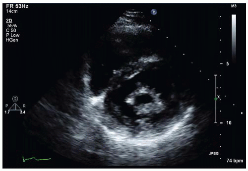 Figure 12-7. |
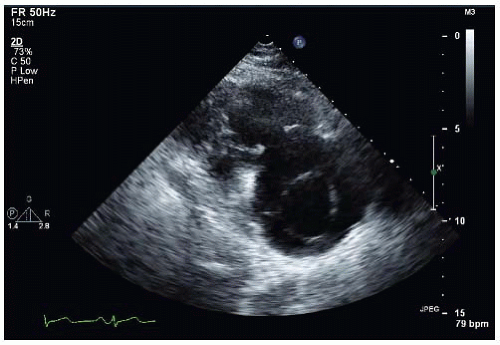 Figure 12-8. |
Stay updated, free articles. Join our Telegram channel

Full access? Get Clinical Tree


