Severe Mitral Regurgitation
A 72-year-old man with severe mitral regurgitation (MR) and a flail P3 scallop undergoes robotic mitral valve surgery. Transesophageal echocardiogram (TEE) images are obtained in the operating room after mitral valve surgery (Fig. 67-1 and Video 67-1).
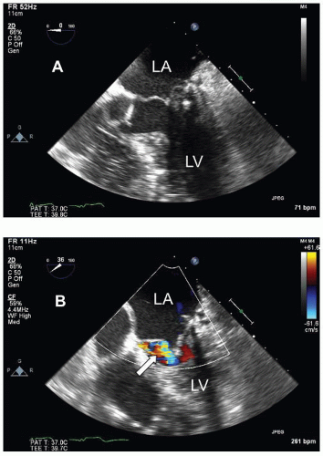 Figure 67-1. A. TEE five-chamber view: End-systole. B. TEE four-chamber view: Diastole. |
QUESTION 1. What type of mitral valve surgery did the patient have?
A. Mitral valve replacement with a bioprosthetic mitral valve
B. Mitral valve repair
C. Mitral valve replacement with a mechanical mitral valve
D. Alfieri stitch
View Answer
ANSWER 1: B. The valve was repaired using a partial annuloplasty ring and triangular resection of a flail P3 segment with closure of the posterior commissure and reattachment of the chords. The valve is still visibly myxomatous. There is no evidence of a bioprosthetic or mechanical valve. The Alfieri stitch would not be used for a P3 prolapse/flail segment.
A. Residual MR
B. Mitral stenosis after the repair
C. Aortic regurgitation
D. Systolic anterior motion (SAM) of the mitral apparatus with left ventricular outflow tract gradient
View Answer
ANSWER 2: C. It is always important to evaluate for residual MR and for the presence of mitral stenosis after repair. The color jet is seen in diastole on the ventricular side of the mitral valve and can therefore not be due to MR. Similarly, SAM is a systolic event and therefore also excluded. The absence of flow convergence on the atrial side excludes a jet due to mitral stenosis. The color flow represents the presence of new aortic regurgitation. Further imaging is required to better characterize the lesion and severity.
QUESTION 3. The following applies to the aortic regurgitation seen in Figure 67-2 and Video 67-2:
Stay updated, free articles. Join our Telegram channel

Full access? Get Clinical Tree


