Segmental Left Ventricular Systolic Function

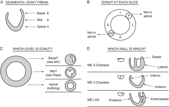
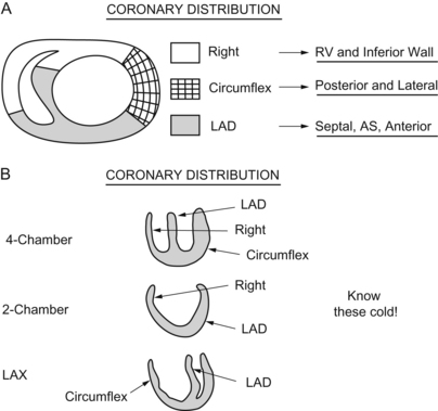
Chapter 14


Segmental Left Ventricular Systolic Function


John C. Sciarra and Christopher J. Gallagher


Myocardial Segment Identification


Hear ye, hear ye. The Office of Homeland Security is not going to shoot me for revealing any state secrets here. You will need to know these segments and you will need to know which coronaries feed which walls and which segments.


I kid thee not.


I speak not with forked tongue.


This is a for sure on the test.


The first time you see it you’ll quasi freak, because it looks so complex, but when you think of all the other stuff you memorized to get this far, it’s not so bad. Plus there’s a logic to it, so don’t go off the deep end.


First, the whole thing, then we’ll back up and break it down.


image


I found it easiest to start with the cross sections. That way you can at least always know what’s “directly across” from you. Then you can take the long views and start to put it together.


So, think of three of these lying on top of each other, starting at the top of the ventricle, right next to the mitral valve. Three layers of six:


The basal 6 segments are next to the mitral valve.


The middle 6 segments are next down, at the level of the papillary muscles.


The lower, or apical, 6 segments come next.


BUT WAIT!


Though it would make sense to do 6/6/6, the SCA (perhaps fretting about the demonic number 666 from The Omen), only recognizes four segments in the ever-narrowing apex.


Lose the posterior and the anteroseptal segments there. In the apical, you just have inferior, anterior (across from each other, remember), and septal and lateral (across from each other too).


Now, put it back together, piece by piece, until it makes sense. If you are still confused, stay tuned for the 17 segment chapter.


Coronary Artery Distribution and Flow


Pictures tell it all:


image


Let’s put it into words, just in case you’re less of a visual learner.


The right coronary feeds the inferior wall and right ventricle.


The left anterior descending feeds the anterior and septal walls. (No wonder an LAD infarct is so problematic.)


The circumflex feeds the posterior and lateral wall.


Everyone studies the hell out of this issue, drawing the pictures over and over again, flashcards, you name it. Get this stuff down but down.


Here is a little memory helper I made up, I call it the “coronary artery memory helper”:


LM → LAD → Diags (the “D” in lad leads into the “D” in diagonals).


Circ → OMs (circumferential looks like the circumference of the “O” in Obtuse Marginal).


RC → PDA (the right hand [RCA] writes on the palm pilot pda).


Jun 4, 2016 | Posted by in CARDIOLOGY | Comments Off on Segmental Left Ventricular Systolic Function

Full access? Get Clinical Tree

Get Clinical Tree app for offline access