Sarcoidosis
Armando E. Fraire MD
Sarcoidosis, a systemic disease of unknown etiology often affecting the lungs and mediastinal lymph nodes, is characterized histopathologically by granulomas that are found distinctively along the path of the bronchovascular bundles. The cardinal diagnostic feature of sarcoidosis in the lung, as in other anatomic sites, is the nonnecrotizing granuloma. This granuloma is made up of tight conglomerates of epithelioid histiocytes and multinucleated giant cells of the Langerhans type, surrounded by a peripheral rim of fibroblasts. Typically, the granulomas are often naked, that is, devoid of a peripheral rim of lymphocytes, a feature that can be seen in nonsarcoidal granulomas. A variety of cytoplasmic inclusions can be seen within the multinucleated giant cells, including asteroid bodies, conchoid bodies, and colorless birefringent particles. None of these inclusions are diagnostic, but their presence may alert the pathologist to the possibility of sarcoid. Rarely, the granulomas may involve the vascular walls or become confluent, leading to conditions known as sarcoid vasculitis or confluent sarcoid, respectively. Transbronchial lung biopsy is very useful in the diagnosis of sarcoidosis, allowing a diagnosis to be made in ≥70% of the cases. However, endobronchial biopsies are also useful. The actual yield of these two diagnostic modalities varies primarily on account of the stage of the disease. The diagnostic yield in either type of biopsy can be increased by step sectioning of the tissue blocks.
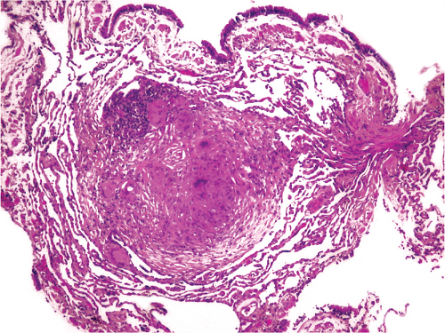 Figure 25.1: A discrete nonnecrotizing granuloma is seen at low power in this transbronchial biopsy from a patient with sarcoidosis.
Stay updated, free articles. Join our Telegram channel
Full access? Get Clinical Tree
 Get Clinical Tree app for offline access

|