CHAPTER 21 Right-Sided Mediastinal Lymph Node Dissection—Video 21
Approach to Video-Assisted Right-Sided Mediastinal Lymph Node Dissection
 Video-Assisted Right-Sided Mediastinal Lymph Node Dissection
Step 1. Level 9 and 8 Lymph Nodes
♦ Aim the thoracoscope anteriorly, and point the 30-degree lens downward toward the inferior pulmonary ligament.
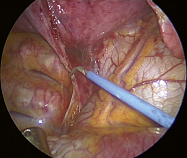
♦ Bring the suction catheter and the long-tipped electrocautery through the anteroinferior incision. Press the curve of the Yankauer suction catheter onto the diaphragm to hold it out of the way.
♦ If the diaphragm is high and in the way, as is seen frequently in obese patients, place a heavy stitch in the ligamentous portion of the diaphragm. Pull the stitch through the incision for the trocar. Suture it to the skin to retract the diaphragm.
♦ Hold the inferior pulmonary ligament toward the apex of the chest. Level 9 lymph nodes can be seen.
♦ Incise the inferior pulmonary ligament with the long-tipped electrocautery up to the level of the inferior pulmonary vein as shown in Figure 21-1. The tip of the suction catheter should be held close to the electrocautery tip to remove the smoke created.
Step 2. Level 7 Lymph Nodes
♦ Aim the thoracoscope anteriorly, and point the 30-degree lens downward toward the inferior pulmonary ligament.
♦ With ring forceps, which are brought through the inferior and the utility incisions, retract the right lower lobe medially toward the heart.
♦ Incise the posterior mediastinal pleura with scissors, which are brought through the anterior incision as shown in Figure 21-2. Carefully incise the pleura at its junction with the lung parenchyma. The natural tendency is to gravitate posteriorly toward the esophagus, but this should be avoided because it causes nuisance bleeding from the esophageal muscle.
Stay updated, free articles. Join our Telegram channel

Full access? Get Clinical Tree


