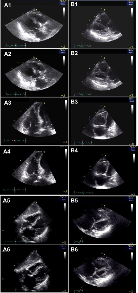
Bedside echocardiography plays an important role in the first-line diagnosis of Takotsubo cardiomyopathy (TC). Several classic imaging features could aid in the differential diagnosis in patients who have manifestation similar to that of acute coronary syndrome and potentially help in the risk stratification and management, including the decision to use coronary angiography. Right ventricular (RV) involvement in TC has been previously identified. However, these abnormal imaging features have never been well characterized and analyzed. Since September 2009, we have studied consecutive cases of TC diagnosed at our institute, with a specific focus on the analysis of RV morphologic abnormalities and their correlation with the unique hemodynamic effects of TC. We present a distinct regional pattern of RV free wall motion that could add additional diagnostic value to the assessment of TC. In patients with TC, the motion of the basilar and middle segments of the RV free wall is often hyperkinetic ( Figure 1 and Videos 1 to 3 ). However, the motion of the apical segment of the RV free wall is usually hypokinetic ( Figure 1 and Video 1 to 3 ), in the same manner as the left ventricular (LV) apical motion ( Figure 2 ). Interestingly, this distinct imaging feature is exactly opposite the classic echocardiographic appearance in patients with acute and massive pulmonary embolism, McConnell’s sign, which is defined as hyperkinesis of the RV apex and hypokinesis of the remaining segments of the RV free wall ( Figure 1 , Video 4 to 6 ).

Stay updated, free articles. Join our Telegram channel

Full access? Get Clinical Tree


