Regional Ventricular Function Assessment
Lori B. Heller1
Solomon Aronson1
Lori B. Heller2
Solomon Aronson2
1OUTLINE AUTHORS
2ORIGINAL CHAPTER AUTHORS
▪ KEY POINTS
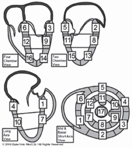
The American Society of Echocardiography (ASE) divides the left ventricle (LV) into 17 segments (Fig. 9-1).
The basal and mid levels of the LV each have inferior, inferior-lateral, anterolateral, anterior, anteroseptal, and inferoseptal segments.
The apical level has inferior, lateral, anterior, and septal segments. The apical cap is the final segment.
All segments can be visualized from either a midesophageal or a transgastric probe location.
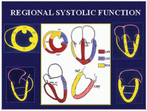
When wall-motion abnormalities are observed, knowing the typical blood supply patterns to these segments can help identify the compromised coronary artery or graft.
A wall-motion description should be assigned for each segment.
Mild hypokinesis refers to decreased (less than 30%) inward wall movement toward the center of the LV during systole (normal being greater than 30%).
Severe hypokinesis refers to only slight wall movement with less than 10% systolic thickening.
Akinesis refers to lack of both movement and endocardial thickening.
Dyskinesis refers to outward wall movement during systole and endocardial thinning during systole.
Since movement may be passive, thickening is felt to be more reliable than endocardial wall motion in determining wall-motion abnormality.
I. SEGMENTAL MODEL OF THE LEFT VENTRICLE
Global and regional ventricular functions are key elements in the evaluation of patients with ischemic heart disease.
Multiple images must be acquired from multiple planes and assumptions must be made about the shape of the ventricle and the coronary artery distribution within the ventricle (Fig. 9-2).
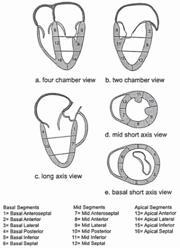
The Society of Cardiovascular Anesthesiologist and ASE have recommended a 16-segment model for regional LV assessment (Fig. 9-3).
American Heart Association (AHA) published a position paper standardizing the segmentation nomenclature based on a 17-segment model with minor differences in nomenclature.
ASE has adopted these new standards; the Society of Cardiovascular Anesthesiologists and the National Board of Echocardiography have not.
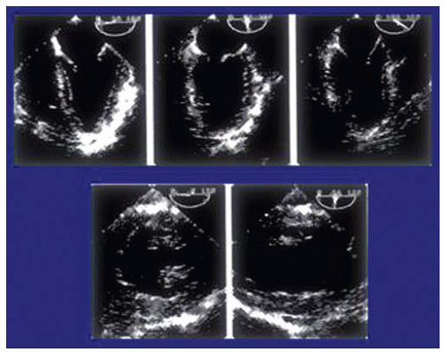
Regional function assessment of the LV can be accomplished by obtaining five standard views (three from the midesophageal window and two from the transgastric window; Fig. 9-4).
Obtaining the midesophageal views of the LV
Position the transducer posterior to the left atrium (LA) at the mid level of the mitral valve.
The imaging plane is then oriented to simultaneously pass through the center of the mitral annulus and the apex of the LV.
The depth should be adjusted to include the entire LV (usually 16 cm).
Rotating to multiplane angle of 0 degree should keep the center of the mitral annulus and LV apex in view.
The midesophageal four-chamber view
Obtained by rotating the multiplane angle forward from 0 degree until the aortic valve is no longer in view and the diameter of the tricuspid annulus is maximized, usually between 10 and 30 degrees.
Shows all three segments (basal, mid, and apical) in each of the septal and lateral walls (Video 9-1; Fig. 9-5).
At the beginning of Video 9-1, the LV outflow tract (LVOT) and part of the aortic valve comes into view. This image should be rotated slightly toward 10 to 20 degrees to obtain a true four-chamber representation (Video 9-1).
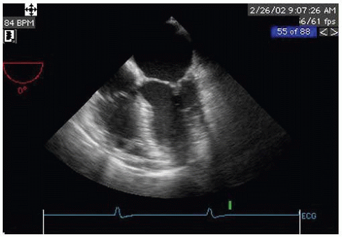
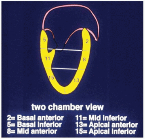
The midesophageal two-chamber view
Obtained by rotating the multiplane angle forward until the right atrium and the right ventricle (RV) disappear, usually between 90 and 110 degrees (Video 9-2).
Shows the three segments (basal, mid, and apical) in each of the anterior and inferior walls (Fig. 9-6).
The midesophageal long-axis view
Developed by rotating the multiplane angle forward until the LVOT, aortic valve, and the proximal ascending aorta come into view, usually between 120 and 160 degrees (Video 9-3).
This view shows the basal and midanteroseptal segments (not the apical segments), as well as the basal and midposterior segments.
Therefore, with the imaging plane properly oriented through the center of the mitral annulus and the LV apex, one can examine the entire LV without moving the probe and simply rotating the multiplane angle from 0 to 180 degrees.
The transgastric views of the LV
Acquired by advancing the probe into the stomach and anteflexing the tip until the heart comes into view.
At multiplane angle of 0 degrees a short axis of the LV should appear.
The probe is then turned to the right or left as needed to center the LV in the display.
The depth should be adjusted to maximize the entire LV, usually 12 cm.

Stay updated, free articles. Join our Telegram channel

Full access? Get Clinical Tree


