Fig. 10.1
Groin anatomy in recurrent varicose vein. Type I. SFJ patent. Type I A main stem (GSV intact). Type I B tributary (missed tributaries in the groin). Type I C neovascularization. FV femoral vein, GSV great saphenous vein
A.
Main stem
B.
Tributary
C.
Neovascularization
Type 2 Recurrent – saphenofemoral venous complex obliterated. This has two subdivisions (Fig. 10.2).
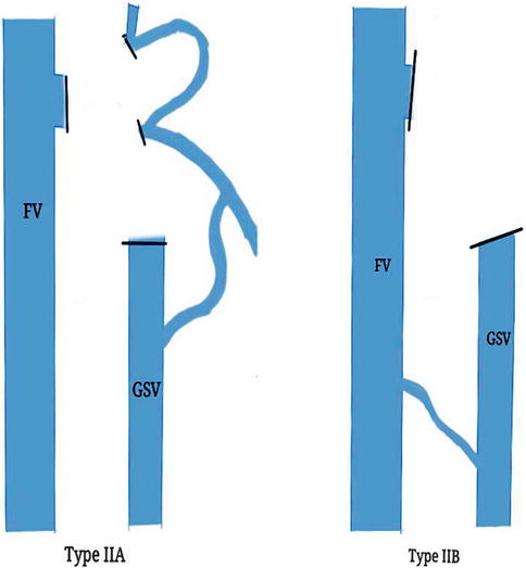
Fig. 10.2
Groin anatomy in recurrent varicose veins – type II. SFJ obliterated. Type II A cross groin. Type II B mid thigh. FV femoral vein, GSV great saphenous vein
A.
Cross groin
B.
Mid thigh
The presence of GSV was noted and the length of thigh vein graded on a 0–3 scale.
0 = no GSV
1 = GSV present in lower thigh
2 = present in upper thigh but not to the groin
3 = present to the groin
The current most widely accepted system of classification for recurrent varicose veins is the REVAS classification. This would be discussed in detail.
REVAS Classification
The REVAS classification includes six items:
Topographic sites of REVAS – T
Source of reflux – S
Degree of reflux – R
Nature of sources – N
Contribution from persistent incompetent saphenous trunk – P
Possible contributory factors – F
1.
Topographic sites (T)
Groin (g) | 1 |
Thigh (t) | 2 |
Popliteal fossa (p) | 3 |
Lower leg including ankle and foot (l) | 4 |
Others (o) | 5 |
2.
Sources of reflux (S)
No identified source | 0 |
Pelvic and/or abdomen | 1 |
SFJ | 2 |
Thigh perforators | 3 |
SPJ | 4 |
Popliteal perforator | 5 |
Gastrocnemius vein | 6 |
Lower leg perforators | 7 |
3.
Degree of reflux (R)
Probable clinical significance | R+ |
Unlikely clinical significance | R− |
Uncertain clinical significance | R? |
4.
Nature of sources (N)
N ss – same site of previous surgery
Technical failure | 1 |
Tactical failure | 2 |
Neovascularization | 3 |
Uncertain/unknown | 4 |
Mixed | 5 |
N ds – different site
Persistent | 1 |
New | 2 |
Uncertain/unknown | 3 |
5.
Contribution from a persistent incompetent saphenous trunk (P)
GSV – AK (above knee) | 1 |
GSV – BK (below knee) | 2 |
SSV | 3 |
Neither/others | 4 |
6.
Possible contributory factors (F)
General factors (Fg)
Family history | 1 |
Obesity | 2 |
Pregnancy | 3 |
OCP | 4 |
Lifestyle factors (prolonged standing, lack of exercise, chair sitting) | 5 |
Specific factors (Fs)
1. Primary deep vein reflux | 1 |
2. Post-thrombotic syndrome | 2 |
3. Iliac vein compression | 3 |
4. Angiodysplasia | 4 |
5. Lymphatic | 5 |
6. Calf pump dysfunction | 6 |
In practice, this system is applied with the aid of the REVAS sheet which contains a checklist for all the parameters. The REVAS classification is much more comprehensive and was found to be useful for routine clinical practice as well as research studies. The intraobserver concordance of the system was found to be satisfactory, but the interobserver concordance was not uniform [1].
Stay updated, free articles. Join our Telegram channel

Full access? Get Clinical Tree


