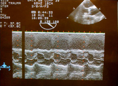
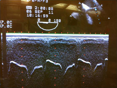
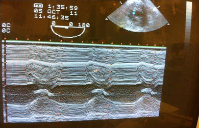
Chapter 6 Quantitative M-mode and Two-dimensional Echocardiography Varun Dixit, John C. Sciarra and Christopher J. Gallagher M-mode provides a one-dimensional, “ice pick” view through the heart and updates the B-mode images at a very high rate.1 Doppler recordings reflect blood velocity, whereas M-mode motion of cardiac structures reflects volumetric blood flow. The two examinations are hemodynamically complementary. In certain situations, M-mode recordings of the valves and inter-ventricular septum can be particularly helpful.2 M-mode is a superior method of examining the timing of the cardiac events especially when displayed with the electrocardiogram. When measuring “where is the real live edge” in the ventricle, the back of the mid-papillary muscle view is the most appropriate. Then, the whole deal centers on where does the blood stop and where does the ventricle start. For this, you are best off using contrast, as noted above. Other than that anemic explanation, I don’t know beans about how they’d ask you a test question on “Edge Components”. Deeper field?—That would take more information in, so temporal resolution would be worse. Shallower field?—That would take less information in, so temporal resolution would be better. Wider field?—More information coming in, therefore worse temporal resolution. Narrower field?—You’re getting it. Less info, therefore better temporal resolution. First you will see the chest wall. The next hypoechoic shadow is the right ventricular outflow tract. The next ‘ice pick’ line you get is the aortic anterior wall. The ‘box car’ represents the cusp separation of the aortic valve. Note, the two leaflets of the mitral valve move in M-shaped mirror image pattern in diastole. At the onset of systole the two leaflets come together and generate the first heart sound.
Edge Components
Temporal Resolution
Aortic Valve in M-Mode

Mitral Valve in M-Mode

Thoracic Key
Fastest Thoracic Insight Engine


