Pulmonary Infections in Patients with Human Immunodeficiency Virus Disease

Chapter 29 Pulmonary Infections in Patients with Human Immunodeficiency Virus Disease


Epidemiology, Risk Factors, and Pathophysiology


Human Immunodeficiency Virus Infection: Background


It is now more than 30 years since the first report from Los Angeles, in the United States, of an “outbreak” of Pneumocystis pneumonia (PCP) among homosexual men that heralded the onset of the global human immunodeficiency virus (HIV) pandemic. Since the early 1980s, HIV infection has steadily spread throughout the world and caused an estimated 25 million deaths. At the end of 2009, according to the United Nations Program on HIV/AIDS (UNAIDS), 33.3 million people worldwide were HIV-infected, a majority of these living in resource-poor countries. In 2008, the Centers for Disease Control and Prevention (CDC) reported that in the United States, 1,178,350 persons 13 years of age or older were living with HIV infection, and more than 40,000 were newly infected each year. In the United Kingdom, the Health Protection Agency estimated that in 2009, 86,500 persons were living with HIV infection and 6630 were newly diagnosed. Over the previous 10 years, the proportion of U.K. residents older than 50 years of age who were living with HIV infection had increased from 1 in 20 to 1 in 5.


Current antiretroviral therapy (ART) regimens have the potential to suppress HIV replication for decades. In 2011, persons with newly diagnosed HIV infection living in nations such as the United States and the United Kingdom that provide access to treatment and care have an expectation that on ART their life expectancy will approach that for an age-matched HIV-uninfected population. Their clinicians now manage HIV-infected populations whose medical complications reflect age-related comorbidity. Unfortunately, these observations do not apply to a majority of people worldwide with HIV infection, who live in financially impoverished environments with little access to comprehensive health care prevention and treatment programs.


Respiratory disease remains an important contributor to morbidity and mortality, and more than two thirds of HIV-infected persons have at least one respiratory episode during the course of their illness. With relatively preserved immune responses, infectious agents are similar to those seen in the general population, although at a higher frequency. With progressive HIV disease, subjects are at an increased risk for opportunistic disease. For example, the North American Prospective Study of Pulmonary Complications of HIV Infection (PCHIS), a multicenter cohort drawn from all HIV risk groups at various stages of immunocompromise, revealed that over an 18-month study period, of approximately 1000 subjects who were not using ART, 33% reported an upper respiratory tract infection, 16% had an episode of acute bronchitis, 5% acute sinusitis, 5% bacterial pneumonia, and 4% developed PCP.


The immune dysregulation that arises from HIV infection means that bacteria, mycobacteria, fungi, viruses, and protozoa can all cause disease in patients with advanced infection. Box 29-1 shows the organisms that typically infect the lung in HIV disease. Of these, the agents of bacterial infections, tuberculosis, and PCP are the most important. In the West, 40% of diagnosed AIDS cases are due to PCP. This chapter provides a brief general overview of the epidemiology and pathogenesis of HIV infection, followed by a more detailed discussion of other important aspects of the disease and its infectious pulmonary complications.


It is reported that by the end of 2009, 33.3 million people worldwide had acquired HIV infection (Figure 29-1). Of these, over 40% are thought to have developed AIDS (for definition of AIDS, see Tables 29-1 and 29-2 and Box 29-2). Globally, 2.6 million people acquired HIV infection in 2009, and 1.8 million died of AIDS. The developing world has been most affected. Sub-Saharan Africa is the current epicenter of the pandemic (accounting for two thirds of all infections); here, nearly 6% of adults are HIV-infected. South and Southeast Asia are responsible for almost a fifth of the estimated HIV global burden. In Central-Eastern Europe and Central Asia, there are currently 1.4 million HIV-infected persons. In the developed world, North America and Western Europe account for approximately 1.5 million and 820,000 infections, respectively. The vast majority of these are spread through sexual contact, although vertical (mother-to-child) and blood-borne infections are common. In the developing world, heterosexual transmission is the norm. In North America and Europe, men who have sex with men constitute the largest group of HIV-infected persons.


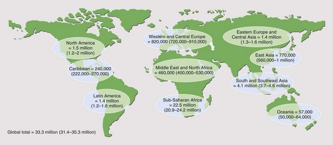
Table 29-1 CDC Classification of HIV Infection


Group Infection
I Acute primary
II Asymptomatic
III Persistent generalized lymphadenopathy
IV Other disease
Subgroup A Constitutional disease (e.g., weight loss >10% of body weight or >4.5 kg; fevers with temperatures >38.5° C for >1 month; diarrhea lasting >1 month)
Subgroup B Neurologic disease (e.g., HIV encephalopathy, myelopathy, peripheral neuropathy)
Subgroup C Secondary infectious diseases
Subgroup C1 AIDS-defining secondary diseases (e.g., Pneumocystis jirovecii pneumonia, cerebral toxoplasmosis, cytomegalovirus retinitis)
Subgroup C2 Other specified secondary infectious diseases (e.g., oral candidiasis, multidermatomal varicella zoster)
Subgroup D Secondary cancers (e.g., Kaposi sarcoma, non-Hodgkin lymphoma)
Subgroup E Other conditions (e.g., lymphoid interstitial pneumonitis)

AIDS, acquired immunodeficiency syndrome; HIV, human immunodeficiency virus.
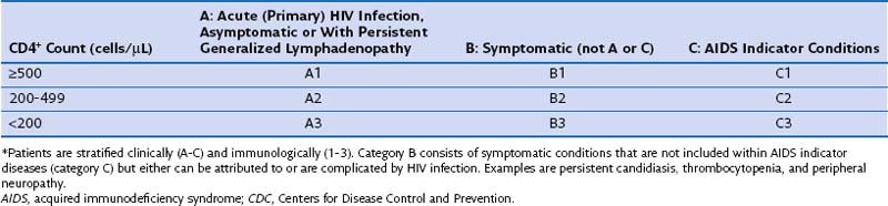


Virology and Immunology of the Human Immunodeficiency Virus


HIV was first isolated in 1983 from patients with symptoms and signs of immune compromise. Two subtypes, HIV-1 and HIV-2, have subsequently been identified. HIV-1 (hereafter referred to as HIV) is responsible for a majority of infections, is associated with a more aggressive clinical course, and is the focus of this chapter.


HIV is a human retrovirus belonging to the lentivirus family. Cell-free or cell-associated HIV infects through attachment of its viral envelope protein (gp120) to the CD4 antigen complex on host cells. The CD4 receptor is found on several cell types, although the T helper lymphocyte is the main site of HIV infection in the body. Additionally, HIV gp120 also must bind to the cell surface protein co-receptor chemokine receptor 5 (CCR5), or to other co-receptors, including CXCR4, depending on the host cell type. Polymorphisms in genes coding for CCR5 may affect disease progression by reducing the ability of HIV to enter and infect cells. At a population level, this effect appears to be small.


Once HIV is inside the cell, it uses the enzyme reverse transcriptase (RNA-dependent DNA polymerase) to transcribe its own RNA into a DNA copy that translocates into the nucleus and integrates with host cell DNA using its viral integrase. The virus (as proviral DNA) remains latent in many cells until the cell itself becomes activated. This may arise from cytokine or antigen stimulation. The viral genetic material is then transcribed into new RNA, which, in the form of newly created virions, bud from the cell surface and infect other host CD4-bearing cells.


HIV infection directly attacks the immune system, and in particular the T helper cells that underpin the coordinated immune response. This leads to progressive immune dysfunction, with an inability to react to opportunistic pathogens as well as persistent, unregulated immune activation. The etiopathogenic process is not well defined, although at the time of primary infection it is thought that HIV spreads to regional lymph nodes, circulating immune cells, and thymus. The result is a massive viral infection of the human host, which, despite a relatively potent immune response, targets specific memory T cells responsible for sustaining long-term protective immunity. Without therapeutic intervention, progressive immune failure is inevitable. This occurs through a combination of direct cell killing caused by HIV replicating within cells and the negative effects of chronic immune activation. Ultimately, in most cases the infection produces immune system destruction and dysfunction, which is reflected in a reduction in circulating absolute blood CD4+ cell count, a decrease in the percentage of T cells expressing CD4 markers, and a fall in the CD4+-to-CD8+ T cell ratio. Patients present with clinical disease indicating profound immunodeficiency.


Natural History of Human Immunodeficiency Virus Infection


Intervention with ART, as well as specific preventive (prophylactic) therapy for opportunistic infections has changed the clinical presentation of HIV disease in countries in which these interventions are available. Death rates for HIV-infected cohorts have fallen precipitously. In the absence of ART, in the developed world the median interval between HIV seroconversion and progression to AIDS is estimated to be 10 years and is shorter in cash-poor countries. Untreated, almost all cases of HIV infection progress to full-blown AIDS; without ART, 95% of affected patients will die within 5 years. Globally, in 2011 the main causes of death among HIV-infected patients (a majority of whom were not receiving ART) included tuberculosis, enteric bacterial infection, bacterial pneumonia, and PCP.


The clinical course of untreated HIV infection evolves in several reasonably distinct stages. First, acquisition of the virus, next, seroconversion, which (in the minority) may be associated with a clinical illness (primary HIV infection), then follows a clinically silent period lasting several months to years which leads to symptoms and signs indicating progressive HIV-induced immune compromise, ultimately resulting in AIDS (e.g., PCP).


Chronic Human Immunodeficiency Virus Infection


Although a proportion of HIV-seropositive persons remain completely well without ART for an extended period (approximately 20%, after 10 years), many infected persons have minor symptoms and signs suggesting immune dysfunction. Oral candidiasis and constitutional symptoms (e.g., malaise, idiopathic fever, night sweats, diarrhea, weight loss) are the strongest clinical predictors of progression to AIDS.


The term AIDS was originally created as an epidemiologic tool to capture specific clinical presentations, which early in the HIV epidemic appeared to suggest significant immune deficiency. Over the past 30 years, the definition has been modified to incorporate the expanding spectrum of diseases affecting HIV-infected patients, including cervical carcinoma and recurrent bacterial pneumonia (see Box 29-2). The 1993 CDC classification included an immunologic criterion for AIDS (CD4+ count below 200 cells/µL or CD4+ percentage less than 14% of total lymphocytes) regardless of clinical symptoms (see Table 29-2). These data are used to define a point at which the risk for severe opportunistic infection rises dramatically.


Apart from cervical carcinoma, AIDS indicator diseases differ little between men and women. Injection drug users in the United States and the United Kingdom have a high incidence of recurrent bacterial pneumonia and tuberculosis. Geographic differences occur that reflect the opportunistic pathogens present in the local environment (e.g., histoplasmosis or visceral leishmaniasis usually are found only in patients from endemic areas). In the developed world, survival differences after an AIDS diagnosis mainly arise from variation in ease of access to and provision of medical care. It is clear that better treatment outcomes are associated with specialist care provided by treatment centers with extensive experience in the management of HIV-infected persons.


In countries in which ART is available, the spectrum of HIV-related disease has changed over the past 30 years. In the HIV Outpatient Study (HOPS), a prospective multicenter observational study in the United States, between 1994 and 2007, opportunistic infections associated with very low CD4+ counts (e.g., cytomegalovirus retinitis, Mycobacterium avium complex [MAC] infection) declined rapidly after introduction of ART and stabilized at low levels during the period 2003 to 2007. In the EuroSIDA cohort (a pan-European prospective study of HIV infection), between 1994 and 2004, opportunistic infections were observed less frequently over time, and malignant disease, such as non-Hodgkin lymphoma, increased as an AIDS-defining event.


Although death rates have fallen in ART-treated populations, there has been a rise in the proportion of non-AIDS deaths. In some series, this category accounts for a majority of events. Causes include liver disease (often due to viral hepatitides) and cancer, as well as cardiovascular disease and drug-related toxicity. In such circumstances, AIDS deaths usually occur among patients who have not accessed medical care regularly and who present with advanced HIV disease.


A new manifestation of opportunistic infection has been described in patients commencing ART. The immune reconstitution inflammatory syndrome (IRIS) (the pathogenesis of which is discussed later under treatment for tuberculosis) may cause severe if temporary clinical illness as the patient’s immunity recovers. Patients appear to experience a relapse of their original (and often incompletely treated) disease. IRIS often is seen in MAC infection, tuberculosis, hepatitis B, CMV retinitis, and herpesvirus infection. In the developed world, metabolic complications of ART, such as ischemic heart disease, hypertension, diabetes, and cerebrovascular disease, are increasingly encountered by clinicians providing care. A significant number of persons receiving ART also experience drug toxicity. An increasing number of patients also are surviving to manifest symptoms associated with chronic hepatitis B and C virus infection. HIV-associated nephropathy (often with chronic kidney disease) is common among black Africans and is a significant cause of long-term morbidity.


Pulmonary Immune Response During Human Immunodeficiency Virus Infection


It is clear from the frequency with which HIV-related respiratory disease occurs that the pulmonary immune response is profoundly compromised. Evidence from simian immunodeficiency virus (SIV)-infected primates indicates that within the lung acute retroviral infection causes a rapid increase, followed by a decline in SIV RNA. Intrapulmonary replication is “compartmentalized,” in that plasma SIV levels correlate poorly with those in bronchoalveolar lavage (BAL) fluid. In humans, comparison of HIV replication in blood and in alveolar lining fluid indicates that little intrapulmonary HIV replication occurs when patients are asymptomatic. This process increases significantly with development of pulmonary disease, and in some instances, local HIV replication is greater than in plasma. Other studies, however, show no consistent change in HIV levels in BAL fluid as the patient’s clinical status alters. Pulmonary memory CD4+ cells are not as extensively infected by HIV compared with those within the gastrointestinal tract. Alveolar macrophages appear to carry a much lower HIV “viral load” than macrophages obtained from other body sites.


HIV affects both the humoral and cellular components of innate immunity. These alterations are apparent even in patients with (near) normal CD4+ counts and undetectable HIV loads, and include a BAL CD8+ lymphocytosis. HIV-induced impairment of innate immune function, particularly when there is additional CD4+ cell depletion, likely contributes to the pathogenesis of opportunistic pulmonary infections, for example, the increased risk of mycobacterium tuberculosis infection and bacterial pneumonia.


Phagocytosis of bacteria by human alveolar macrophages does not appear to be influenced by HIV infection, suggesting that other mechanisms are important. In the general population deficiencies in signaling through Toll-like receptors (TLRs) are associated with an increased risk of, or an adverse outcome from, a number of infections. Activation of HIV-infected human macrophages by TLR4 results in impaired tumor necrosis factor-α release compared to that from uninfected macrophages; as interleukin (IL)-10 release is not impaired, this effect appears specific.


Intra-pulmonary immune responses after ART initiation are not as well described as those occurring during untreated HIV infection. Detection of HIV in BAL fluid is less likely in subjects on ART. Starting such therapy is associated with a delayed but significant decrease in the absolute number and percentage of alveolar CD8+ lymphocytes. Successful virologic control leads to a reduction in activated intrapulmonary CD8+ cells and increases in CD8+-naive and central memory cells—implying that the intrapulmonary CD8+ lymphocyte pool is repopulated from the peripheral circulation.


ART also induces a marked reduction in concentrations of proinflammatory cytokines and chemokines in BAL fluid. Despite this, both IFN-γ and IFN-γ inducible chemokines (inducible protein [IP]-10, monokine induced by IFN-γ [MIG]) remain detectable and appear to contribute to recruitment of memory cells into the lung.


On the basis of these findings, it is hypothesized that ART-naive patients have uncontrolled intrapulmonary HIV replication, which results in nonspecific cellular activation and augmented cytokine and chemokine expression. These changes promote an influx of inflammatory cells into the alveoli. With institution of ART, intrapulmonary HIV load and cellular activation are reduced and nonspecific cytokine secretion resolves, although persistent, low-level IFN-γ production from resident memory cells maintains IFN-γ–inducible chemokine levels. As a result, normal intrapulmonary trafficking of these cells occurs, resulting in more clear-cut evidence of innate and acquired immune control. The former includes ART reducing mononuclear cell chemokine production with a subsequent impact on TLR2-mediated signaling.


Risk Factors for Respiratory Disease


An individual patient’s risk for respiratory disease is determined by the medical history (e.g., receipt of effective preventive therapy or ART), place of residence and travel history (e.g., the influence of geography on mycobacterial and fungal disease), and immunity status. Falling blood CD4+ counts or high plasma HIV RNA “viral loads” increase the chance of respiratory infection, with an increased spectrum of potential organisms associated with greater degrees of immunosuppression. For example, HIV-infected patients with a CD4+ count below 200 cells/µL are four times more likely to have one episode of bacterial pneumonia per year than those with higher CD4+ cell counts. More exotic organisms are found in subjects with very low CD4+ counts. These include bacteria such as Rhodococcus equi and Nocardia asteroides and fungi such as Aspergillus spp. and Penicillium marneffei. Just as with P. jirovecii, this increased susceptibility reflects the importance of T cell depletion and macrophage dysfunction in the loss of host immunity (a process that has been confirmed by animal experiments).


Among HIV-infected patients, injection drug users are at greatest risk for development of bacterial pneumonia and tuberculosis. Persons who have had previous respiratory episodes (PCP or bacterial pneumonia) appear to be at increased risk for subsequent episodes of such disease. Whether this susceptibility relates to host or environmental factors is not certain, although structural lung damage and abnormal pulmonary physiology are likely to be contributing factors. This argument is supported by the increased rates of pneumonia in HIV-infected smokers compared with nonsmokers. Recent work has shown that chronic obstructive pulmonary disease (COPD) and lung cancer occur more frequently and at a younger age among HIV-infected patients, than in the general population. In view of the large number of HIV-infected persons who smoke heavily, targeting this population for smoking cessation obviously is a pressing need. This goal is reinforced by the association between smoking and an increased risk for bacterial pneumonia and more rapid progression to first AIDS illness and death.


Clinical Features


Bacterial Infection


Bronchiectasis


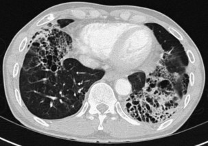
Bronchiectasis is increasingly recognized in HIV-infected patients with advanced HIV disease and low CD4+ lymphocyte counts. It probably arises secondary to recurrent bacterial, mycobacterial or P. jirovecii infections. The diagnosis most often is made by high-resolution (thin-section) computed tomography (CT) scanning (Figure 29-2). Its prevalence has not been accurately determined, although with improved survival from both opportunistic infections and HIV disease, it can be expected to be increasingly common in clinical practice. The pathogens isolated in patients with bronchiectasis are those seen in bronchitis. In addition, Burkholderia cepacia and Moraxella catarrhalis have been described.


Pneumonia


Community-acquired bacterial pneumonia occurs more frequently in HIV-infected patients than in the general population. It is especially common in HIV-infected injecting drug users. The spectrum of bacterial pathogens is similar to that in non–HIV-infected persons (see Box 29-1). S. pneumoniae is the most commonly identified pathogen, followed by H. influenzae. HIV-infected patients with S. pneumoniae–related pneumonia frequently are bacteremic. In one study, the rate of pneumococcal bacteremia in HIV-infected patients was 100 times that for an HIV-negative population. More recent work has confirmed this to be the case for all causes of HIV-related bacterial pneumonia. Typically, blood cultures have a 40-fold increased pick-up rate in HIV-positive patients. The widespread use of ART has led to some decrease in rates of bacterial pneumonia and bacteremia, although they are still considerably higher than in a non–HIV-infected population.


Bacterial pneumonia has a similar presentation in HIV-infected patients and in uninfected persons. Chest radiographs frequently are atypical in appearance, mimicking that in PCP in up to half of the cases (Figure 29-3). By contrast, radiographic lobar or segmental consolidation also may be seen in a wide range of bacterial organisms (Figure 29-4); these include S. pneumoniae, P. aeruginosa, H. influenzae, and M. tuberculosis. PCP also may manifest with lobar or segmental consolidation. In patients with more advanced HIV disease and low CD4+ lymphocyte counts, P. aeruginosa and S. aureus also can cause pneumonia.
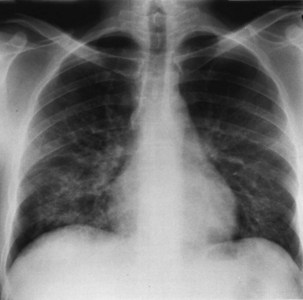
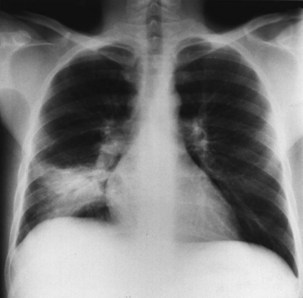


Complications of bacterial pneumonia are frequent, and pleural effusions are twice as likely in HIV infection (often occurring with S. aureus infection); empyema and intrapulmonary abscessation are present in up to 10% of patients. Inevitably, the mortality rate is high (approximately 10%).


Other Bacterial Infections


Mycobacterial Infections


Tuberculosis


HIV infection is associated with a 20- to 40-fold increased risk for development of active tuberculosis disease in a person with latent tuberculosis infection (LTBI) over that in noninfected subjects. Taken together with its ability to infect both the immunocompromised and the immunocompetent, tuberculosis is perhaps therefore the single most important disease associated with HIV infection. It is estimated that each year, 1.1 million new cases of active tuberculosis occur in HIV-infected patients. Accordingly, tuberculosis is a major cause of HIV-related morbidity and mortality. It also is a clear driver in both resource-rich and poor countries for the current overall increase in tuberculosis rates. Where HIV infection is endemic, tuberculosis control at a population level is almost impossible if treatment for both infections is not available.


In the United Kingdom, many centers routinely offer HIV antibody testing to all patients with tuberculosis, regardless of risk factors for HIV infection. In the United States, the CDC now recommends HIV testing as a routine part of health care for all patients 13 to 64 years of age seeking medical services. The benefit of treating HIV co-infection is indisputable. Moreover, strategies that reduce high-risk behavior and cut ongoing HIV transmission can be introduced. Unfortunately, offering HIV testing in tuberculosis clinics has not been routine practice in many countries, and integration with HIV infection–related services is poor. This disconnect leads to both diagnostic delay of HIV infection and potentially suboptimal, uncoordinated care, with potentially disastrous outcomes.


Active tuberculosis can occur at any stage of HIV infection and, unlike almost every other HIV-related infection, may do so despite effective ART. In the United States, the United Kingdom, and most European countries, reporting of tuberculosis in both HIV-infected and non–HIV-infected patients is mandatory.


Clinical disease in HIV-infected patients may arise in several different ways: by reactivation of latent tuberculosis, by rapid progression of pulmonary infection, and by reinfection from an exogenous source.


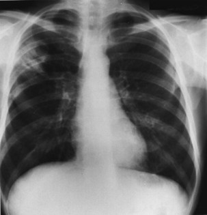
Pulmonary disease is the most common presentation, and clinical manifestations are determined by the patient’s level of immunity. For example, persons with reasonably well-preserved CD4+ counts exhibit clinical features similar to those of “normal” adult postprimary disease (Table 29-3). Signs and symptoms typically include weight loss, fever with sweats, cough, sputum, dyspnea, hemoptysis, and chest pain. These patients may have no clinical features to suggest associated HIV infection. The chest radiograph frequently shows upper lobe consolidation, and cavitary change is common (Figure 29-5). If performed, the tuberculin skin test (TST), using purified protein derivative (PPD), usually gives a positive result, and the likelihood that spontaneously expectorated sputum or BAL fluid will be smear-positive for acid-fast bacilli is high.


Table 29-3 Tuberculosis and Human Immunodeficiency Virus (HIV) Infection


Diagnostic Feature Stage of HIV Disease
Reasonable Immunity Impaired Immunity
Chest radiographic appearance Upper zone infiltrates and cavities (cf postprimary infection) Lymphadenopathy, effusions, miliary or diffuse infiltrates (cf primary infection)
Normal
Sputum or bronchoalveolar lavage “smear-positive” Frequently Less commonly
Disease site Localized Widely disseminated
Tuberculin test–positive Frequently Less commonly


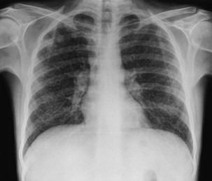
In persons with advanced HIV disease (i.e., low CD4+ lymphocyte counts and clinically apparent immunosuppression), it may be difficult to diagnose tuberculosis. The clinical presentation here often is with nonspecific symptoms. Fever, weight loss, fatigue, and malaise may be mistakenly ascribed to HIV infection itself. In this context, pulmonary tuberculosis is often similar to primary infection, with the chest radiograph showing diffuse or miliary-type shadowing (Figure 29-6), hilar or mediastinal lymphadenopathy, or pleural effusion; cavitation is unusual, with no upper zone chest radiographic predominance. In up to 10% of patients, the chest radiograph may appear normal; in others, the pulmonary infiltrate can be bilateral, diffuse, and interstitial in pattern, thus mimicking PCP. Hilar lymphadenopathy and pleural effusion also may be manifestations of pulmonary Kaposi sarcoma or lymphoma, with which M. tuberculosis may coexist. The TST result usually is negative, and spontaneously expectorated sputum and BAL fluid samples often are smear-negative (although they are culture-positive).


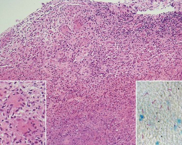
In addition to pulmonary tuberculosis, extrapulmonary disease occurs in a high proportion of HIV-infected persons with low CD4+ lymphocyte counts (less than 150 cells/µL). Mycobacteremia and generalized lymph node infection (Figure 29-7) are common, but involvement of bone marrow, liver, pericardium, meninges, and brain also has been described.


Evidence of extrapulmonary tuberculosis should be sought in any HIV-infected patient with suspected or confirmed pulmonary tuberculosis, by culture of stool, urine, and blood or bone marrow. Traditional solid phase culture and speciation techniques may take 6 to 10 weeks. Liquid culture methods (e.g., BACTEC, Becton Dickinson, Towson, Maryland) that detect early growth provide a faster diagnosis (usually in 2 to 3 weeks). Molecular diagnostic tests using M. tuberculosis genome detection (e.g., by polymerase chain reaction [PCR]) offer the possibility of yet more rapid diagnosis (within hours). The original concerns that “high-tech” systems may not be practical in a field setting are now being challenged. The World Health Organization (WHO) has endorsed the Xpert TBRIF assay, which is portable and simple to use with sputum samples. This kit is expensive, however, although it may be of use in high-prevalence tuberculosis/HIV infection settings, in which prompt treatment initiation is critical to infection control. Current data suggest that such tests are of value, although less sensitive in HIV-infected subjects (in whom sputum samples generally have a lower bacterial load). The availability of simple but highly sensitive and specific methods that utilize the inoculation of large quantities of the sample (e.g., sputum) onto microscopic plates with subsequent rapid detection (in days) of both mycobacterial growth and resistance patterns (i.e., microscopic observation drug susceptibility [MODS] assay) is of great potential significance.


Until the results of culture and speciation are known, acid-fast bacilli identified in respiratory samples, biopsy tissue, an aspirate, or blood in an HIV-infected patient, regardless of the CD4+ lymphocyte count, should be regarded as being M. tuberculosis, and conventional antituberculosis therapy should be commenced. If culture fails to demonstrate M. tuberculosis and instead another mycobacterium (see later) is identified, then the treatment regimen can be modified.


Drug-Resistant Tuberculosis


Multiple drug–resistant (MDR) tuberculosis—that is, disease caused by M. tuberculosis strains resistant to isoniazid and rifampicin (rifampin), with or without other drugs—is now an important clinical problem in HIV-infected patients in the United States, where it is responsible for approximately 3% of all cases of tuberculosis in this population. Outbreaks of MDR tuberculosis have occurred in both HIV-infected and non–HIV-infected persons in the United States in prison facilities, hostels, and hospitals. Similar incidents also have been documented among HIV-infected patients in Europe. Inadequate treatment (including case management and supervision of medication) of tuberculosis and poor patient compliance with antituberculosis therapy are the most important risk factors for development of MDR tuberculosis. Other cases have arisen through exogenous reinfection of profoundly immunosuppressed HIV-infected patients who are already receiving treatment for drug-sensitive disease.


Despite antituberculosis therapy, the median survival in HIV-infected persons with MDR tuberculosis initially was only 2 to 3 months. Recently this has improved, largely because of an increased awareness of the condition with early initiation of suitable therapy as determined by drug sensitivity testing.


The advances in molecular diagnostics are not confined to rapid mycobacterial speciation. Commercial kits are available which can detect the commonest mutations that confer rifampicin and isoniazid resistance with good sensitivity. These are of great potential value in infection control in tuberculosis-endemic areas. They can guide initial therapy, assess for resistance in unexpected poor clinical response and significantly improve treatment outcomes. Next generation tests will also incorporate drug resistance profiles for other important agents such as fluoroquinolones.


Infections Due to Mycobacteria Other Than Tuberculosis


Pneumocystis jirovecii Pneumonia


The development of PCP is largely related to underlying states of immunosuppression induced by malignancy or treatment thereof, organ transplantation, or HIV infection. In 2011 in the United States, the United Kingdom, Europe, and Australasia, PCP was largely seen only in HIV-infected persons unaware of their serostatus or in those reported to be intolerant of or noncompliant with anti–P. jirovecii prophylaxis and ART.


P. jirovecii originally was regarded as a protozoan, as suggested by its morphology and the lack of response to antifungal agents such as amphotericin B. The organism is now thought to be a fungus. The demonstration of antibodies against P. jirovecii in most healthy children and adults suggests that infection is acquired in childhood and persists in the lungs in a dormant phase. Subsequent immunosuppression (e.g., as a result of HIV infection) allows the fungus to propagate in the lung, causing clinical disease. This “latency hypothesis,” however, is challenged by several observations: P. jirovecii cannot be identified in the lungs of immunocompetent persons; “case clusters” of PCP in health care facilities suggest recent transmission; different genotypes of P. jirovecii are identified in each episode in HIV-infected patients who have recurrent PCP; genotypes of P. jirovecii in patients who have PCP correlate with place of diagnosis and not with their place of birth, suggesting infection has been recently acquired. Taken together, these data suggest that PCP arises by reinfection from an exogenous source.


The clinical presentation of PCP is nonspecific, with onset of progressive exertional dyspnea over days or weeks, together with a dry cough, with or without expectoration of minimal quantities of mucoid sputum. Patients often report an inability to take a deep breath, which is not due to pleurisy (Table 29-4). Fever is common, yet patients rarely complain of temperature-related signs and symptoms including sweats. In HIV-infected patients, the presentation usually is more insidious than in those receiving immunosuppressive therapy. The median time to diagnosis from onset of symptoms is more than 3 weeks in those with HIV infection, compared with less than 1 week in non–HIV-infected patients. In a small proportion of HIV-positive patients, the disease course of PCP is fulminant, with an interval of only 5 to 7 days between onset of symptoms and progression to development of respiratory failure. In others, it may be much more indolent, with respiratory symptoms that worsen almost imperceptibly over several months. Rarely, PCP may manifest as a fever of undetermined origin without respiratory symptoms.


Table 29-4 Clinical Presentation in Pneumocystis jirovecii Pneumonia


Examination Typical Presentation Atypical Presentation
Symptoms Progressive exertional dyspnea over days or weeks Sudden onset of dyspnea over hours or days
Dry cough ± mucoid sputum Cough productive of purulent sputum
Hemoptysis
Difficulty taking in a deep breath not related to pleuritic pain Chest pain (pleuritic or “crushing”)
Fever ± sweats
Tachypnea
 
Signs Normal breath sounds or fine end-inspiratory basal crackles Wheeze, signs of focal consolidation or pleural effusion
Chest radiographic appearance Early: perihilar “haze,” or bilateral interstitial shadowing Pleural effusion, lobar or segmental consolidation
Late: alveolar-interstitial changes or “whiteout” (marked alveolar consolidation with sparing of apices and costophrenic angles)
Arterial blood gases PaO2: early: normal; late: low  
PaCO2: early: normal or low; late: normal or high

Clinical examination usually is remarkable only for the absence of physical signs; occasionally, fine, basal, end-inspiratory crackles are audible. Features that would suggest an alternative diagnosis include a cough productive of purulent sputum or hemoptysis, chest pain (particularly pleural pain), and signs of focal consolidation or pleural effusion (see Table 29-4). Of note, infection with more than one pathogen occurs in almost one fifth of these patients, so symptoms may be related to infection with any of several agents.


The chest radiographic appearance in PCP typically is unremarkable initially. Later, diffuse reticular shadowing, especially in the perihilar regions, is seen and may progress to widespread alveolar consolidation that resembles that in untreated pulmonary edema or with presentation late in disease. At this stage, the lung may be grossly consolidated and almost airless (Figure 29-8). Up to 20% of chest radiographs are atypical in appearance, showing lobar consolidation, honeycomb lung, multiple thin-walled cystic air spaces (pneumatoceles), intrapulmonary nodules, cavitary lesions, pneumothorax, and hilar and mediastinal lymphadenopathy. Predominantly apical changes, resembling those of tuberculosis, may occur in patients with PCP that developed subsequent to anti–P. jirovecii prophylaxis with nebulized pentamidine (Figure 29-9

Only gold members can continue reading. Log In or Register to continue

Stay updated, free articles. Join our Telegram channel

Jun 12, 2016 | Posted by in RESPIRATORY | Comments Off on Pulmonary Infections in Patients with Human Immunodeficiency Virus Disease

Full access? Get Clinical Tree

Get Clinical Tree app for offline access