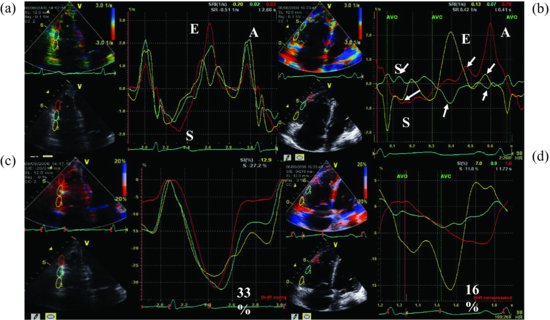
RV dysfunction was further confirmed by tissue Doppler image analysis (Figure 25.2).
Figure 25.2 The longitudinal strain rate of right ventricular segments from a normal person (a). The longitudinal strain rate is decreased and desynchronized in a patient with pulmonary embolism (b). The peak of longitudinal segment strain is 33% and synchronized from a normal person (c). The peak of longitudinal segment strain is decreased and desynchronized (d) in our case.
Initial electrocardiogram showed sinus tachycardia with very mild ST elevation in leads V1 and V2.
Stay updated, free articles. Join our Telegram channel

Full access? Get Clinical Tree


