Never-smokers (n = 1,178)
Active smokers (n = 777)
p
Smoking status (pack-years)
0
30.0 (15.0–43.2)
Age (year)
65.3 ± 10.1
56.2 ± 10.3
<0.001
Male Gender (%)
45.4
77.9
<0.001
BMI
27.4 ± 4.2
27.0 ± 4.2
0.833a
LDL-C (mg/dl)
119.1 ± 36.4
117.5 ± 32.1
0.012a
HDL-C (mg/dl)
41.2 ± 11.1
36.2 ± 10.2
0.002a
Triglycerides (mg/dl)
136 (102–192)
154 (112–218)
<0.001c
Preexisting CVD (%)
68.1
80.1
<0.001b
Diabetes mellitus (%)
38.3
36
0.314b
Hypertension (%)
76.6
63.3
<0.001b
Lipid lowering drugs (%)
42.4
52.8
<0.001
hsCRP (ng/ml)
2.7 (1.2–7.0)
4.9 (1.8–10.3)
<0.001c
LpPLA2 (ng/ml)
383.8 (272.3–533.5)
424.2 (293.7–630.2)
<0.001c
Smoking status in the group of active smokers showed a large variability, ranging from 0.3 up to 132 pack-years. Males were more frequent in the group of active smokers and the mean age was lower. Plasma concentrations of LDL-C and HDL-C were significantly higher in never-smokers, whereas total triglycerides were higher in active smokers. However, results for LDL-C were likely affected by treatment with lipid lowering drugs (mainly statins), which was more frequent in active smokers. Preexisting CVD was also more prevalent in active smokers, whereas a higher prevalence of arterial hypertension was observed in never-smokers. However, body mass index (BMI) and prevalence of diabetes mellitus were inappreciably different between the study groups (Table 1).
Valid measurements of both hsCRP and LpPLA2 were available for 1,048 never-smokers and 685 smokers. Plasma concentrations of hsCRP and LpPLA2 were significantly higher in active smokers than in never-smokers (hsCRP, 4.9 (1.8–10.3) ng/ml vs. 2.7 (1.2–7.0) ng/ml; LpPLA2, 424.2 ng/ml (293.7–630.2) ng/ml vs. 383.8 (272.3–533.5) ng/ml, respectively; p < 0.001 for both comparisons) (Table 1).
As a next step we analyzed the proportion of concentrations higher or lower than the median value of hsCRP and LpPLA2 in either study group. Three hundred forty (49.6 %) of active smokers had plasma concentration of hsCRP and 343 (50.0 %) had LpPLA2 above the median. Concentrations above the median for both markers were found in 190 (27.7 %) and below the median in 192 (28.0 %) of active smokers. Five hundred eighteen (49.4 %) of never-smokers had plasma concentrations of hsCRP and 523 (49.9 %) had LpPLA2 above the median. Concentrations above the median for both markers were found in 277 (26.4 %) and below the median in 284 (27.1 %) of never-smokers (Table 2).
Table 2
Absolute and relative number of patients with concentrations of hsCRP and LpPLA2 higher or lower than the median
Never-smokers | Active smokers | |
|---|---|---|
1,048 | 685 | |
hsCRP low, LpPLA2 low | 284 (27.1 %) | 192 (28.0 %) |
hsCRP low, LpPLA2 high | 246 (23.5 %) | 153 (22.3 %) |
hsCRP high, LpPLA2 low | 241 (23.0 %) | 150 (21.9 %) |
hsCRP high, LpPLA2 high | 277 (26.4 %) | 190 (27.7 %) |
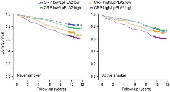
Within the observation time (median of 10 years), 995 patients died. From these, 221 were active smokers (28.4 % out of the 777) and 302 never-smokers (25.6 % out of the 1,178); the difference between active smokers and never-smokers did not reach here significance (log rank test p = 0.212). The Kaplan-Meier curves were calculated for both groups depending on the concentrations of hsCRP and LpPLA2 below or above the median as described above. In active smokers, the subgroup with the concentration of both markers above the median showed the worst survival compared with all other groups (Fig. 1). The survival difference in the different subgroups is compiled in Table 3.


Fig. 1
Survival of patients with values of hsCRP and LpPLA2 higher or lower than the median
Table 3
Dependence of survival on concentrations of hsCRP and LpPLA2 higher or lower than the median
Never-smokers (NS) | Active smokers (S) | p (χ2 test) | |||
|---|---|---|---|---|---|
n patients | n events | n patients | n events | NS vs. S | |
1,048 | 264 (25 %) | 685 | 197 (29 %) | 0.100 | |
hsCRP low, LpPLA2 low | 284 | 44 (15 %) | 192 | 48 (25 %) | 0.010 |
hsCRP low, LpPLA2 high | 246 | 54 (22 %) | 153 | 40 (26 %) | 0.337 |
hsCRP high, LpPLA2 low | 241 | 68 (28 %) | 150 | 41 (27 %) | 0.850 |
hsCRP high, LpPLA2 high | 277 | 98 (35 %) | 190 | 68 (36 %) | 0.927 |
p (log-rank test) | <0.001 | <0.02 | |||
In never-smokers, a stepwise decrease of survival was observed in patients with both hsCRP and LpPLA2 below the median, hsCRP below and LpPLA2 above the median, hsCRP above and LpPLA2 below the median, and hsCRP and LpPLA2 above the median; with the best survival in cases with a low concentration and the worst in those with a high concentration of both markers (Fig. 1, Table 3).
Comparison of mortality rates of active smokers and never-smokers demonstrated a large similarity in all groups, except for the group with low concentration of both markers. This group displayed a significant difference between active smokers and never-smokers, with a χ2 p-value of 0.010 (Table 3).
To investigate whether the observed differences in survival between the smokers and never-smokers, according to the concentration of hsCRP and LpPLA2, were influenced by a different distribution of risk factors between both groups, we conducted the Cox regression analysis with adjustment for age and sex (Model 1) or with an additional adjustment for diabetes, hypertension, coronary artery disease, BMI, LDL-C, HDL-C, and triglycerides (Model 2). For the analysis, we divided smokers and never-smokers according to tertiles of hsCRP and LpPLA2 concentrations and compared the groups with both markers in the lowest or highest tertile, and hsCRP and LpPLA2 each in the highest tertile. For both smokers and never-smokers, there was a significant increase in risk for patients in whom the concentration of both hsCRP and LpPLA2 was in the upper tertile. When only one marker was elevated, there was a trend toward increased risk, which became significant only in Model 2 in never-smokers (Table 4).


Table 4
Cox regression of survival dependence on tertiles of hsCRP and LpPLA2 concentrations adjusted for risk factors
< div class='tao-gold-member'>
Only gold members can continue reading. Log In or Register to continue
Stay updated, free articles. Join our Telegram channel

Full access? Get Clinical Tree


