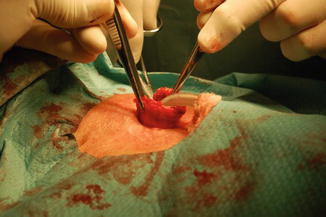
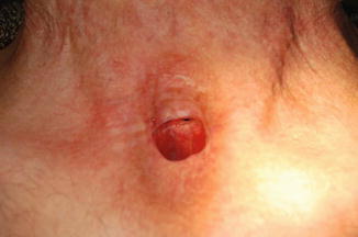
Fig. 1
Tracheoesophageal (T-E) shunt decay at day 6 after secondary TE puncture

Fig. 2
Mobilization and resection of trachea with reconstruction of esophagus

Fig. 3
Persistent treacheoesophageal fistula after reconstruction
In April of 2012, the patient underwent the Witzel gastrostomy. The swallowing difficulties developed 6 months later. The esophageal X-ray contrast passage revealed a stenosis at the hypopharyngeal junction. In November of 2012, flexible gastroscopy confirmed the presence of stenosis without mucosal fold in the upper third of the esophagus. Direct repeat esophagoscopy uncovered circular stenosis with ∅ 2 mm approximately 4 cm above the tracheostomy. The stenosis was gradually dilated up to the probe No. 32. Because of inflammation and edema at the site of stenosis, dilation was discontinued and parenteral anti-inflammatory and anti-edematous treatment was initiated. The patient complained of dyspepsia caused by delay in gastric emptying. He lost 23 kg of body weight between April and November of 2012. The patient’s condition improved after introduction of gastric prokinetics. He was finally released in stable condition for outpatient care. Further re-dilation of the stenosis was performed in December of 2012 up to the probe No. 34, after which the swallowing of solid food significantly improved. However, a leak of liquids persisted. A rarely diagnosed material intolerance was suggested as a source of the patient’s problems.
3 Discussion
The prosthetic voice rehabilitation developed after a failure of laryngectomized patients to use esophageal voice or electrolarynx (Singer and Blom 1980). The basic condition of successful voice restoration in these patients is an adequate opening pressure in the pharyngoesophageal segment. It seems that anatomical conditions are more important than the patient’s mental need to talk (Sebova-Sedenkova 2010).
The insertion of a tracheoesophageal prosthesis may be followed by different adverse effects. A minor consequence is tissue granulation around the puncture site (Imre et al. 2013). Treatment includes antibiotics, antifungals, chemical or electrocautery, and surgical excision of the granulation tissue. Another common complication is biofilms formation. Voice prostheses are usually made of silicon rubber. A continuous exposure to saliva, food, drinks, and oropharyngeal microflora contributes to the rapid colonization of the prosthesis by biofilms consisting of mixed bacteria and yeast strains leading to failure and frequent replacement (Talpaert et al. 2014). Microbial colonization and biofilm formation can lead to salivary leakage through voice prosthesis and deterioration of the prosthesis due to the blocking of a valve mechanism. Valve failure as well as compromised speech may result in aspiration pneumonia, and repeat valve replacement may lead to the tract stenosis or insufficiency. Prevention and control of biofilm formation will therefore be beneficial not only for the life span of the prosthesis, but also for the general patient’s health. A number of different approaches have been suggested to inhibit or minimize biofilm formation (for review see Talpaert et al. 2014).
A circumferential enlargement of the tracheoesophageal puncture is a challenging complication as it results in a leakage around the voice prosthesis into the airway and may eventually lead to aspiration pneumonia and respiratory complications (Mobashir et al. 2014). Several treatment alternatives have been proposed to manage the enlarged tracheoesophageal puncture, with varying success. Surgical options include a submucosal purse-string suture around the enlarged tracheoesophageal puncture and its complete closure. Conservative methods, such as customization of the tracheoesophageal voice prosthesis (Lewin et al. 2012), temporary removal of the voice prosthesis (VP) to facilitate stenosis of the tracheoesophageal tract and tracheoesophageal puncture site injections have often been preferred over surgery (Shuaib et al. 2012).
Imre et al. (2013) have retrospectively analyzed 47 patients with secondary tracheoesophageal prosthesis. Tracheoesophageal puncture and speech valve related complications were observed in 20 patients. Minor complications included granulation tissue formation (2 patients), deglutition of prosthesis (6 patients) and tracheoesophageal puncture enlargement/leakage around the prosthesis (9 patients). Major complications were observed in three patients. Two patients developed life-threatening complications: a mediastinitis and paraesophageal abscess, both in the first month of the postoperative period. The overall complication rate was 42.6 % during a mean follow-up of 15.3 months. Tracheoesophageal fistula enlargement was the most common minor complication and the most common cause of complete closure of tracheoesophageal puncture in that study.
Another retrospective study analyzed 103 patients who underwent total laryngectomy or pharyngolaryngectomy (Bozec et al. 2010). Functional outcomes were recorded 6 months postoperatively. A total of 87 patients (84 %) underwent tracheoesophageal puncture and speech valve placement (79 primary and 8 secondary punctures). A high level of comorbidity was correlated to speech rehabilitation failure. Minor leakage around the valve occurred in 26 % of patients. Late complications occurred in 14 patients, including severe enlargement of the fistula (3 patients), prosthesis displacement (7 patients), and granulation tissue-formation (4 patients).
In the present study, out of the 31 patients with voice restoration by secondary tracheoesophageal puncture, 25 (80.6 %) developed inflammation and 13 (41.9 %) developed granulation, which are considered mild complications. No mediastinitis, bleeding, or prosthesis deglutition were recorded.
Despite short-term complications, the prostheses are considered to be well-tolerated in the long-term view. In a cohort of 100 patients (Lukinović et al. 2012), rehabilitation was successful in 75.8 % of patients. The early complication rate was 4.4 %, and 10.9 % of patients had late complications. Statistical analysis failed to substantiate any differences regarding the complication rate and success rate of rehabilitation between the groups of patients stratified according to age, irradiation status, or timing of prosthesis insertion.
< div class='tao-gold-member'>
Only gold members can continue reading. Log In or Register to continue
Stay updated, free articles. Join our Telegram channel

Full access? Get Clinical Tree


