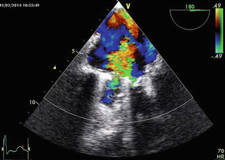
Fig. 17.1
Transesophageal echocardiography showing two physiologic jets due to lavage volume

Fig. 17.2
Transesophageal echocardiography showing the perivalvular leakage due to dehiscence of the prosthesis
Final Diagnosis
Acute heart failure caused by severe mitral regurgitation due to dehiscence of the prosthetic valve. Hemolytic anemia due to the mechanical disruption of red blood cells.
The patient has been recommended to undergo further surgery, but he refused and asked to be discharged. After 3 weeks, he experienced the same symptoms and was admitted again at the hospital. Since the early clinical decompensation, the patient agreed with the previous plan. He underwent a successful mechanical valve replacement.
The patient was keen to switch from warfarin to one of the new oral anticoagulants (NOACs). Unfortunately, while NOACs were proved to be more effective and safer than vitamin K antagonist in prevention of stroke in patient with non-valvular atrial fibrillation, they failed in the setting of valvular atrial fibrillation especially in patients with mechanical prosthetic valves and severe mitral stenosis. Warfarin is, at the present time the only available anticoagulant for patients with prosthetic valvular atrial fibrillation. Since that, no switch was possible for the patient.
17.2 Prosthetic Valve Dysfunctions
Prosthetic valve dysfunctions may be due to regurgitations or obstructions. Sometimes, in case of high velocity flow rate, it is possible to encounter hemolytic anemia due to the mechanical disruption of red blood cells. It ranges from a mild level to a critical hemolysis requiring blood transfusions. This happens mainly in perivalvular regurgitations, but also in normal or accelerated anterograde flow in a mechanical valve.
There are several types of regurgitations that must be differentiated. The most important classification is between pathologic and physiologic regurgitations [1–5].
Mechanical Prosthetic Valves
In mechanical prosthetic valves, color Doppler is the mainstay in identifying, localizing, and grading the severity of regurgitation. The knowledge of the specific model and type of the prosthetic valve we are facing is mandatory. At the same time, it’s important to keep in mind that regurgitation of a prosthetic valve can be physiologic. There are two different types of physiologic regurgitations: The first is the closure volume, that is, the volume of blood pulled back by the closure of the occluders and is confined just after the valve closure; the second is the lavage volume that consists in the blood flow that passes between the occluders and the prosthetic ring throughout the closing state of the valve and is functional to the correct washing of the valve elements. A caged ball valve realizes small and variable jets along the whole circumference of the ball. A tilting disk valve presents jets, one at each side of the occluder; in the bileaflet valve, we can see three regurgitant jets: one central and two near the hinges; making a distinction from perivalvular leakages could be very difficult [4].
Pathologic intra-valvular regurgitation could be secondary to the blockage of an occluder in opening position due to thrombosis, vegetation, or pannus, that is, a fibrous overgrowth of the endocardial tissue over the valve elements. When facing a perivalvular or suspected perivalvular regurgitation, we are called to distinguish a lavage volume passing near the hinges of the occluders from an anomalous regurgitation due to an initial prosthetic valve detachment. Pathologic perivalvular regurgitations are bigger than physiologic ones and show an extensive aliasing. Grading criteria are the same of regurgitation in native valves although the eccentricity and the valve masking phenomenon could make a purely quantitative approach misleading [6].
Biologic Prosthetic Valve
Regarding biologic prosthetic valve, physiologic regurgitation rarely occurs 2 years before the implant, but when present has a central origin, a short length, and low velocities without aliasing. It is important to bear in mind that biologic stentless valves could present physiologic central regurgitation also just after the implant. Pathologic regurgitations of different origin, direction, and nature, either intra- or perivalvular, can occur due to calcific degeneration and endocarditis that could damage a biologic valve even more frequently than a native one.
Prosthetic valve detachment is the most dramatic complication that should be monitored after the recognition of a true perivalvular regurgitation. It is commonly due to an infective endocarditis, frequently with an early onset within 1 year from the implant and related to the implant itself. The infective process disrupts the sewing ring directly generating a perivalvular regurgitation or creating a periprosthetic aneurysm that could be silent until its emptying, when a loss of tissue occurs and the valve anchorage is lost. Beside regurgitation that could range from moderate to severe leading to heart failure manifestations, we can see the rocking movement of the valve that is a sign of a more extensive detachment with a more urgent indication to surgery. The most dreadful consequence is prosthetic valve embolization.
On the opposite side, we can face prosthetic valve obstruction generating valve stenosis of different severity [3]. In this setting, knowing the exact type (mechanical, single disk, bileaflet or biologic, stented/stentless, etc.), model, and dimension of the valve is of paramount importance because the normal values reported by the manufacturers differ in relation to these factors. Moreover, it is essential, at every follow-up, to know the early post-implant gradient values in order to avoid an incorrect diagnosis of prosthetic valve dysfunction. In mechanical valves, the obstruction could be suspected in the presence of a gradient higher than expected for the specific prosthesis. Sometimes the cause is evident such as the presence of a voluminous mass (thrombus or vegetation) occupying space and then reducing the effective orifice area or blocking an occluder in close position; sometimes the cause consists of a small pathologic process that interferes with the hinges that is not directly visible with 2D echocardiography. In those cases, we must look for an anomalous or incomplete opening of the occluders, although it is often difficult to see without the use of fluoroscopy.
< div class='tao-gold-member'>
Only gold members can continue reading. Log In or Register to continue
Stay updated, free articles. Join our Telegram channel

Full access? Get Clinical Tree


