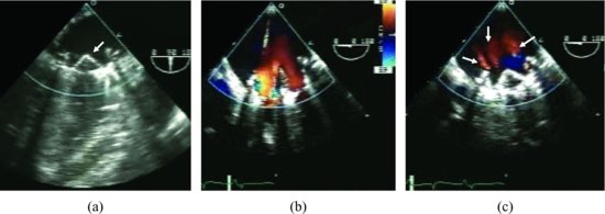
Figure 15.2 Normal St. Jude mechanical mitral valve prosthesis: Its two discs (arrow) are well seen (a). Color flow image of a normal St. Jude mechanical mitral valve in diastole shows acceleration of flow across the valve with two large lateral orifices and a small central orifice (b). Three normal “wash” jets (arrows) are well seen in systole (c).
Transthoracic echocardiography is usually sufficient for evaluation of an aortic prosthesis, but in the mitral position the atria are shadowed by a mechanical prosthesis. Transesophageal echocardiography (TEE), which images from a position posterior to the heart, allows the mitral valve to be seen. TTE demonstrates a ventricular side of the valve that lies in its near field, where as TEE does not. Both methods may have difficulty with the evaluation of the aortic prosthesis. Both echocardiographic methods should be used since they are complementary.
Prosthetic valvular regurgitation
Stay updated, free articles. Join our Telegram channel

Full access? Get Clinical Tree


