Prosthetic Heart Valves


Prosthetic Heart Valves


Allen P. Burke, M.D.


Background

Starr and Edwards described the first successful prosthetic valve replacement in 1961.1 The ball-and-cage mechanical prosthesis was replaced by the (single) tilting disc valve in the 1970s and then, in the 1980s, by the St. Jude Medical bileaflet prosthesis. Although the durability of mechanical valves is superior, the risk of thromboembolic complications and bleeding related to anticoagulation has led to the widespread use of bioprosthetic valves, including porcine valves, homografts, and valves constructed from bovine pericardium.


Types of Prosthetic Valves

Just over half of implanted valves are mechanical, and of these, most are of the bileaflet tilting disc type.

Porcine aortic valve bioprostheses have been the most commonly implanted tissue valves, although bioprosthetic valves fabricated from xenograft pericardium, especially bovine, are becoming increasingly popular. Bioprosthetic valves are usually stented, but may lack a rigid structure and use, for example, porcine aorta as the valve ring.


TABLE 178.1 Types of Prosthetic Valves


Bioprosthetic


Xenograft


Animal valves, either intact aortic porcine valve or valves constructed from three separate semilunar fragments of bovine pericardium


Stented (sewing ring)


Valve or valve fragments attached to mechanical ring


Porcine valve


Intact porcine aortic valve, native, sewed onto metallic ring


Pericardial valve


Fashioned from three separate fragments of bovine pericardium, attached to sewing ring


Unstented


Portion of aorta and porcine aortic valve sewn into tubular cloth graft without sewing ring


Homograft


Human valves


Allograft


Donor’s beating heart or cryopreserved human valves


Autograft


Patient’s own tissue


Pulmonic valve


Transfer of patient’s pulmonic valve to aortic position in congenital aortic valve disease


Transcatheter bioprosthetic


Aortic


Tissue valve mounted on expandable stent


Pulmonary


Tissue valve mounted on expandable stent


Mechanical


Ball cage


Obsolete


Tilting disc


Single disc


Bileaflet


Most common design in use today


Bioprosthetic valves are usually inserted during an open surgical procedure that may be minimally invasive or be inserted via a transcatheter approach, in the case of degenerative trileaflet aortic stenosis. Self- or balloon-expanding valves, in addition to transcatheter insertions, have been designed for surgical valve replacements, for ease of insertion with minimal if any suturing.


Tissue Valves

Tissue valves are either xenografts or homografts (Table 178.1).

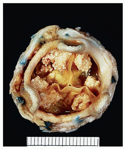
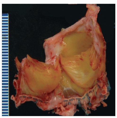
Xenografts are made from preserved animal tissues that are usually mounted on a fabric-covered prosthetic frame or stent (Figs. 178.1, 178.2, 178.3). There are two common types, intact porcine aortic valves and valves constructed from bovine or porcine pericardium. The frame consists of posts (struts) and the intervening valve ring. Stentless xenografts are porcine aortic valves without a rigid stent and often have a portion of porcine aorta attached (full root valves) or lack porcine aorta (subcoronary valves).2

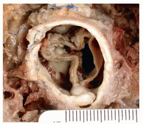
Pericardial xenograft bioprostheses are made of bovine parietal pericardium that is produced by computer-aided design to simulate valve leaflets (Figs. 178.4, 178.5, 178.6).

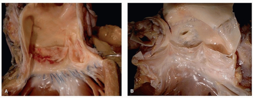
Homografts are human tissue grafts, generally cryopreserved allografts harvested from cadavers or from live donors. These are also stentless, or transplanted directly into the aortic root, without a supporting synthetic frame (Fig. 178.7). Autografts are homografts derived from the patient, usually the pulmonic valve translocated to the aortic position in cases of congenital aortic stenosis.


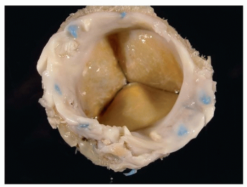
FIGURE 178.1 ▲ Bioprosthetic valve, porcine, removed surgically. The valve shows no evidence of degeneration. One leaflet is open.


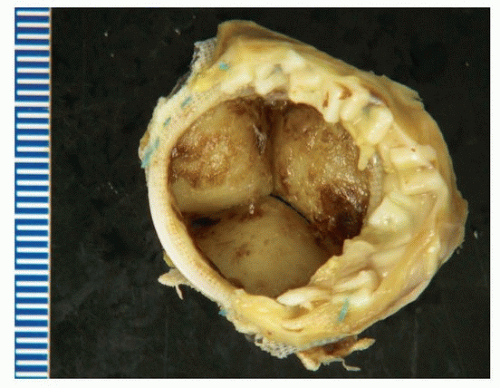
FIGURE 178.2 ▲ Calcific degenerative changes, bioprosthetic porcine valve, necessitating valve replacement. In this case, the calcifications are present as bulky nodules on the leaflet surfaces.


FIGURE 178.3 ▲ Unstented porcine valve, aortic position, removed surgically for degeneration. Two of the porcine leaflets are evident, as well as the cloth sewing ring (bottom), and a portion of the aortic root (above).


FIGURE 178.4 ▲ Pericardial valves. Replacement for excessive pannus overgrowth. In this valve, there is a fibrotic reaction encasing the sewing ring sutures. This response resulted in a degree of stenosis necessitating valve replacement.


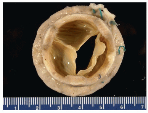
FIGURE 178.5 ▲ Pericardial valve, explant at autopsy. This example has a low profile (Sorin Mitroflow) without metal stent. The cause of death was not related to the valve. The flow surface and sewing ring are shown. There is a small amount of fibrin on the leaflets.


FIGURE 178.6 ▲ Infectious endocarditis, bovine pericardial valve. Two of the three synthetic leaflets demonstrate vegetations.

A list of the more common bioprosthetic valves and their characteristics are presented in Table 178.2.

Mechanical Valves

Bileaflet mechanical valves have largely replaced models of single tilting disc valves (Fig. 178.8). Because of symmetric flow, they offer excellent hemodynamic performance. Most use pyrolytic carbon as the structural component of the leaflets. The current types of bileaflet mechanical valves are presented in Table 178.3.


FIGURE 178.7 ▲ Homograft valve, allograft. Tissue valves may be harvested from human tissue, usually cryopreserved from autopsy. In this example, a human aortic valve was grafted into the pulmonary position in a child with aortic stenosis. A. The native pulmonary valve was normal and was used for an autograft. B. The native pulmonary valve that was removed in from the heart illustrated in Figure 178.9 was moved over into the aortic position, with reimplantation of the coronary ostia (note sutures).


TABLE 178.2 Types of Bioprosthetic Valves, Selected


Class


Examples


Porcine stented


Supra-annular aortic porcine bioprosthesis, Carpentier-Edwards


Duraflex mitral prosthesis, Carpentier-Edwards


Hancock II, Mosaic, Medtronic


St. Jude Medical Biocor


Duraflex


Porcine stentless (full root includes part of pig aorta; subcoronary without root)(aortic position only)


Prima Plus (Carpentier-Edwards)


Freestyle (Medtronic)


SPV Stentless (St. Jude Medical Toronto)


Pericardial stented


Perimount Magna (Carpentier-Edwards)


Theon, Mitral Ease (Carpentier-Edwards)


St. Jude-Biocor


Pericarbon More (Sorin Biomedica)


Pericardial stentless


Freedom Solo, Solo Smart (aortic), Mitroflow (Mitral)(Sorin Biomedica)


Pericardial self expanding (sutureless)


3f Enable (aortic position)(Medtronic)

Only gold members can continue reading. Log In or Register to continue

Stay updated, free articles. Join our Telegram channel

Aug 19, 2016 | Posted by in CARDIOLOGY | Comments Off on Prosthetic Heart Valves

Full access? Get Clinical Tree

Get Clinical Tree app for offline access