(1)
Department of Medicine and Radiology, Stony Brook University Hospital, Stony Brook, NY, USA
This chapter is a compilation of some of the unique and interesting case presentations, which will emphasize on the utilization of cardiac computed tomographic angiography when dealing with procedural complications.
Case 1
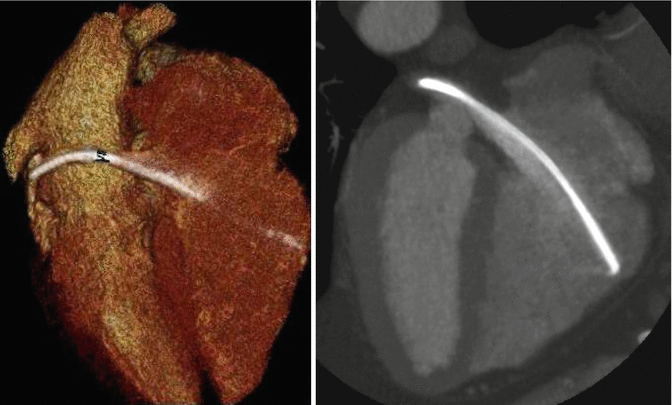
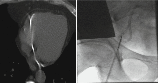
A 55-year-old asymptomatic patient was referred to our emergency department after a routine left subclavian venous port preremoval chest radiograph demonstrated a fractured fragment of the indwelling catheter in the left apical region. A nongated chest CT without contrast failed to precisely reveal location of the embolized fractured distal fragment in the right heart. A transthoracic echocardiogram failed to demonstrate the catheter fragment. An initial attempt to percutaneously remove the embolized fractured fragment using a snare catheter was unsuccessful. A prospectively triggered EKG-gated cardiac CTA was performed, which revealed the fractured catheter fragment lodged in the coronary sinus. The body of the catheter traversed across the tricuspid valve with the tip of the other end abutting the RV free wall. With the precise location of the entrapped catheter identified, the fragment was easily removed by looping a pigtail catheter around the mid portion of the catheter. The superb spatial and temporal resolution of EKG-gated cardiac CTA greatly facilitates preprocedural planning.


Case 2
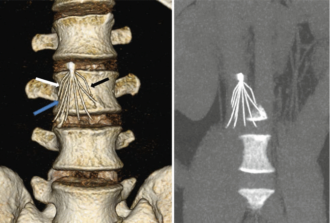
A 32-year-old man with a history of remote right femur fracture presented to the emergency department complaining of dyspnea and sharp chest pain. Initial vital signs demonstrated tachycardia with 104 bpm. CT pulmonary angiogram and transthoracic echocardiogram demonstrated findings suggestive of hemopericardium with tamponade physiology, which was confirmed and treated with pericardiocentesis. Subsequent EKG-gated cardiac CT revealed three identical 2.3 cm linear metallic objects in the following locations: (1) perforating and lodged in the right ventricle free wall (Black arrow), (2) right ventricular cavity (Blue Arrow), and (3) right lower lobe (White arrow). Further patient questioning revealed a history of irretrievable inferior vena cava filter placement at the time of his femur fracture 10 years ago. An abdominal CT revealed an IVC filter missing three of its “arms (Blue, white and black arrows).” The patient was subsequently managed without surgical intervention and is being closely monitored.
 < div class='tao-gold-member'>


Only gold members can continue reading. Log In or Register to continue
Stay updated, free articles. Join our Telegram channel

Full access? Get Clinical Tree


