Recording and Pacing
To discuss intracardiac recording and pacing, we need to introduce two terms that are used by electrophysiologists relating to time measurements. Time measurements are reported in milliseconds (msec, one thousandth of a second), the basic unit of time in electrophysiology.
Cycle Length
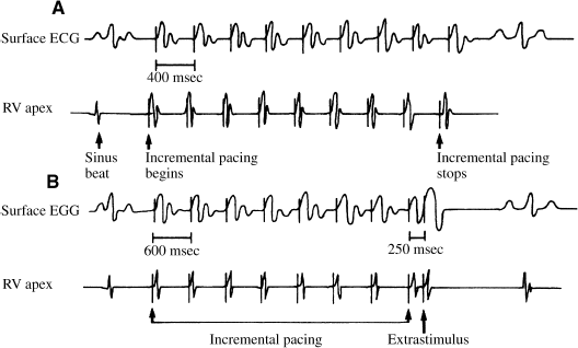
When electrophysiologists talk about heart rate, they typically speak in terms of cycle length—the length of time between each heartbeat (Figure 4.1a). Thus, the faster the heart rate, the shorter the cycle length: an arrhythmia with a rate of 100 beats/min has a cycle length of 600 msec, while an arrhythmia with a rate of 300 beats/min has a cycle length of 200 msec. Non-electrophysiologists must pay close attention when discussing heart rate with electrophysiologists, to avoid some amusing miscommunications.
Figure 4.1 Time measurements in the electrophysiology laboratory. This figure depicts pacing from the right ventricular apex. (a) Cycle length is the interval of time between heartbeats during either incremental pacing (as in this figure) or a spontaneous rhythm. The cycle length of the incrementally paced beats in (a) is 600 msec. (b) Coupling interval is the interval of time between the last normal beat and a premature impulse. In this figure, the normal beats are represented by eight incrementally paced beats at a cycle length of 600 msec. Following the last incrementally paced beat, a single premature stimulus is delivered with a coupling interval of 250 msec.
Coupling Interval
When using a pacemaker to introduce a premature impulse, the coupling interval is the time between the last normal impulse and the premature impulse (Figure 4.1b). With earlier premature impulses, the coupling interval is shorter; with later premature impulses, the coupling interval is longer.
The Electrode Catheter
The electrophysiology study is performed by inserting electrode catheters into blood vessels and positioning these catheters in strategic locations within the heart. Once in position, the catheters can be used to perform the two essential tasks of the electrophysiology study: recording the heart’s electrical signals and pacing.
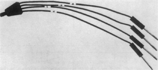
Electrode catheters consist of insulated wires; at the distal tip of the catheter (the end inserted into the heart (Figure 4.2)), each wire is attached to an electrode, which is exposed to the intracardiac surface. At the proximal end of the catheter (the part not inserted into the body (Figure 4.3)), each wire is attached to a plug, which can be connected to an external device (such as a recording device or an external pacemaker). Apart from the fact that they usually have more than two electrodes, these catheters are similar to many temporary pacemaker catheters used in emergency rooms and coronary care units. The catheter shown in Figures 4.2 and 4.3 is a quadripolar electrode catheter, the type most commonly used in electrophysiology studies.
Figure 4.2 The tip of a quadripolar electrode catheter. The electrodes are numbered 1 through 4, the distal (tip) electrode being number 1. The spacing between electrodes is 1 cm.
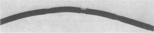
Figure 4.3 The proximal end of a quadripolar electrode catheter. The number on each plug corresponds to the appropriate electrode at the tip of the catheter.
Recording Intracardiac Electrograms
The recording made of the cardiac electrical activity from an electrode catheter placed in the heart is called an intracardiac electrogram. The intracardiac electrogram is essentially an ECG recorded from within the heart. The major difference between a body-surface ECG and an intracardiac electrogram is that the surface ECG gives a summation of the electrical activity of the entire heart, whereas the intracardiac electrogram records only the electrical activity of a localized area of the heart—the cardiac tissue located near the electrodes of the electrode catheter. In general, the intracardiac electrogram records the electrical activity between two of the electrodes (i.e. an electrode pair) at the tip of the catheter. This is called a bipolar recording configuration.
The intracardiac electrogram is filtered electronically, so that in general only the rapid depolarization phase of the cardiac tissue (corresponding to phase 0 of the action potential) is recorded. Figure 4.4 shows an intracardiac electrogram recorded from a catheter in the right atrium during normal sinus rhythm. As the wave of depolarization spreading outward from the SA node passes by the catheter, a discrete high-frequency, high-amplitude signal is recorded. This signal indicates the precise moment at which myocardial depolarization occurs at the electrode pair which is being used for recording. By having catheters positioned in several different intracardiac locations, one can accurately measure the conduction time from one location to another. Further, if enough catheter positions are used, one can map the sequence of myocardial depolarization as an electrical impulse traverses the heart.
Figure 4.4 Surface ECG and intracardiac electrogram recorded from the high right atrium (RA) during sinus rhythm. The deflection recorded on the RA electrogram reflects the precise moment at which the portion of the right atrium at the tip of the electrode catheter is being depolarized.
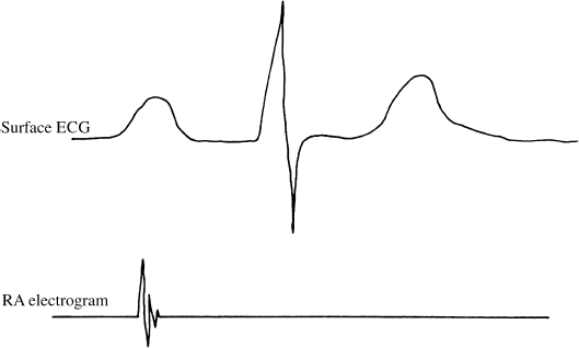
In summary, the deflection recorded from the electrode catheter represents depolarization of the cardiac tissue in the immediate vicinity of the catheter’s electrodes. Thus, the intracardiac electrogram gives precise, localized data on the heart’s electrical impulse.
Pacing
The electrode catheter is also used for pacing. To pace, a pulse of electrical current is carried by the electrode catheter from an external pacemaker to the intracardiac surface, where it causes cardiac cells near the catheter’s electrodes to depolarize. The depolarization of these cardiac cells is then propagated across the heart, just as an electrical impulse arising in the SA node is propagated. Thus, to pace is to artificially generate a cardiac impulse. By careful positioning of the electrode catheter, one can initiate electrical impulses from almost any desired intracardiac location.
During the electrophysiology study, pacing is used to introduce premature electrical impulses, delivered in predetermined patterns and at precisely timed intervals. Such pacing is called programmed stimulation.
There are several reasons for performing programmed stimulation. Precisely timed premature impulses allow us to measure the refractory periods of cardiac tissue. By introducing premature impulses in one location and recording electrograms in other locations, one can assess the conduction properties of the intervening cardiac tissue and the pattern of myocardial activation. Programmed stimulation can also help to assess the automaticity of an automatic focus and to study the presence and characteristics of reentrant circuits.
Programmed stimulation consists of two general types of pacing: incremental and extrastimulus (Figure 4.5).
Figure 4.5 Extrastimulus and incremental pacing. Right ventricular pacing is depicted. (a) Extrastimulus pacing involves introducing one or more extrastimuli either during the patient’s spontaneous rhythm or (as depicted here) following a train of impulses delivered at a fixed cycle length. This panel shows a single extrastimulus. The fixed-cycle-length impulses from which the extrastimuli are timed are referred to as the S1 beats. The first extrasimulus is labeled S2. (b) Same as in (a), except that a second extrastimulus is delivered (labeled S3). (c) Same as in (a) and (b), except that a third extrastimulus is delivered (labeled S4). (d) Incremental pacing consists of a train of paced impulses at a fixed cycle length. The S1 beats in (a) through (c) are incrementally paced impulses.
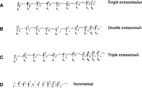
Incremental pacing (or burst pacing) consists of introducing a train of paced impulses at a fixed cycle length. The incremental train may last for a few beats or for several minutes.
The extrastimulus technique consists of introducing one or more premature impulses (called extrastimuli), each at its own specific coupling interval. The first extrastimulus is introduced at a coupling interval timed either from an intrinsic cardiac impulse or from the last of a short train of incrementally paced impulses. (This train is usually eight beats in duration, owing to tradition rather than to scientific reasons.) The generally accepted nomenclature for the extrastimulus technique is illustrated in Figure 4.5. The term “S1” (stimulus 1) is used for the incrementally paced impulses or the intrinsic beat from which the first extrastimulus is timed; “S2” is used for the first programmed extrastimulus; “S3” stands for the second programmed extrastimulus; and so on. This nomenclature (in which, for example, the number 2 is attached to the first extrastimulus) causes a lot of confusion among the uninitiated.
Performance of the Electrophysiology Study
The Physical Setup of the Electrophysiology Laboratory
The electrophysiology study is a type of heart catheterization, and much of the equipment necessary for electrophysiologic testing can be found in a general cardiac catheterization laboratory. This includes a fluoroscopic unit, a radiographic table, a physiologic recorder, oscilloscopes, instrumentation for gaining vascular access, and emergency equipment. Equipment required specifically for electrophysiologic testing includes a programmable stimulator, a multichannel lead switching box (a junction box), and electrode catheters. An arrangement for a typical electrophysiology laboratory is shown in Figure 4.6.
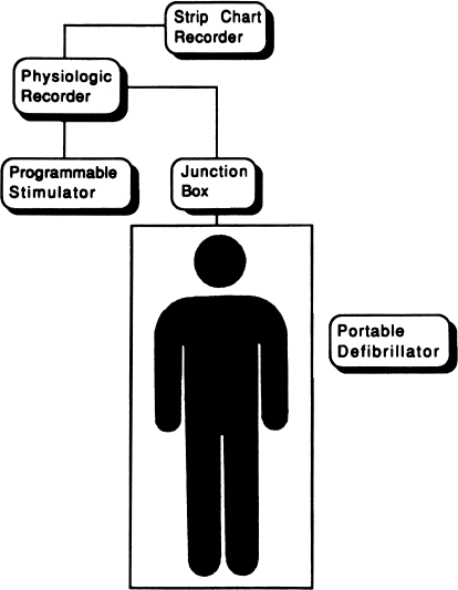
The programmable stimulator is a specialized pacing unit built especially for electrophysiology studies. It has the capability to introduce complex sequences of paced beats with an accuracy of within 1 msec. It can also synchronize pacing to the intrinsic heart rhythm and can pace multiple intracardiac sites simultaneously.
The junction box allows the laboratory personnel to control the connections from the electrode catheters to the various recording and pacing devices. With a series of switches, multiple electrode pairs from multiple catheters can be sorted out for recording and pacing.
The physiologic recorder should have enough channels to process three or four surface ECG leads and multiple intracardiac leads (often up to 12). Most electrophysiology laboratories today are equipped with specialized physiologic recorders designed specifically for electrophysiology studies. Such recorders are computer-based and provide features such as color-coded intracardiac leads, electronic calipers for precise measurements, and hard-disk storage of the entire study.
Equipment to deal with cardiac emergencies is essential, especially because many electrophysiology studies include the intentional induction of arrhythmias, which can cause hemodynamic collapse. This includes a defibrillator, a full complement of cardiac medications, and equipment for maintaining airway support. Likewise, enough trained personnel should always be present to deal with any cardiac emergency. As a minimum, this should include the electrophysiologist, two cardiac nurses who are trained in electrophysiologic procedures (or one nurse and a second physician), and a technician.
Preparation of the Patient
Although preparation of the patient will vary somewhat depending on the nature of the study to be performed, all patients having electrophysiology studies should be given a clear understanding of the purpose and nature of the procedure and the potential benefits and risks. As with any procedure in medicine, the patient should sign a statement of informed consent prior to the study.
Ideally, the electrophysiologic evaluation should be performed while the patient is in a baseline state—non-essential drugs should be withdrawn (especially antiarrhythmic agents), any cardiac ischemia or heart failure maximally treated, electrolytes controlled, and every effort made to prevent excessive anxiety, which can cause excessive sympathetic tone. Anxiety is most easily kept to an acceptable level by adequately preparing the patient for what to expect in the electrophysiology laboratory.
Any cardiac catheterization procedure can cause potentially dangerous arrhythmias, but patients having electrophysiology studies need to be especially aware of this possibility. In particular, patients who are having studies for known or suspected ventricular tachyarrhythmias should be psychologically prepared for the possibility that induction of an arrhythmia might produce loss of consciousness and require defibrillation. Fortunately, the efficacy of electrophysiologic testing and the remarkable safety record achieved in most laboratories go a long way toward allaying the fears of most patients and their families. Detailed discussions with the patient serve not only to guarantee truly informed consent but also to alleviate the patient’s anxiety and build his or her confidence in the electrophysiologist.
Insertion and Positioning of Electrode Catheters
The patient is brought to the catheterization laboratory in the fasting state, and the catheterization sites are prepared and draped. The majority of electrophysiology studies can be performed from the venous side of the vascular system, thus precluding the necessity of catheterizing the arterial tree. Under sterile conditions and after local anesthesia is given, catheters are inserted in most instances percutaneously by the modified Seldinger technique (a needle-stick technique that does not require a cutdown). Only extremely rarely do patients need to be placed under general anesthesia to perform an electrophysiology study; however, premedication with a benzodiazepine is sometimes used in patients who are extremely anxious. Most often, catheters are inserted into the femoral veins (two catheters can safely be inserted into the same femoral vein). Catheters may be inserted from the upper extremities for one of several reasons: for more complicated studies, which require multiple catheters; when there is a contraindication to the use of the femoral veins; when a catheter will be left in place at the end of the procedure; or when positioning of the catheter is easier from the upper extremities (such as in coronary sinus catheterization). In these cases, the internal or external jugular veins, the subclavian veins, or the brachial veins may be used. In those instances where access to the left ventricle is required, the femoral-arterial approach is used most often. In many laboratories, a small catheter is routinely inserted into an artery for continuous monitoring of blood pressure.
Under fluoroscopic guidance, the catheters are placed into the proper intracardiac positions. For a simple diagnostic study, generally one catheter is positioned in the high right atrium and the second catheter is placed in the His position (described later). One of these catheters can later be moved to the right ventricle if ventricular pacing is required. For studies of supraventricular tachycardias, additional catheters are commonly placed in the right ventricle and the coronary sinus (thus allowing recordings from all four major cardiac chambers and from the His position).
In the right atrium, catheters are most commonly positioned in the high lateral wall, near the junction of the superior vena cava. This position approximates the location of the SA node and is the region of the atrium that is depolarized earliest during normal sinus rhythm. Pacing from this area results in P wave configurations that are similar to normal sinus beats.
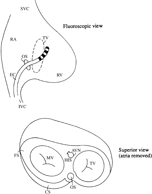
Pacing and recording from the left atrium are usually accomplished by inserting an electrode catheter into the coronary sinus (Figure 4.7). The os of the coronary sinus lies posterior and slightly inferior to the tricuspid valve and is most easily entered from a superior approach (i.e. from the upper extremities). The coronary sinus itself lies in the AV groove—that is, between the left atrium and the left ventricle. Thus, deflections from both the left atrium and the left ventricle are easily recorded from a catheter positioned in the coronary sinus. Pacing the left atrium from the coronary sinus is accomplished routinely—only rarely can the left ventricle be paced from this position. Although a patent foramen ovale will sometimes allow direct entry into the left atrium, we prefer to use the coronary sinus to avoid the slight possibility of causing an embolus in the systemic circulation.
Figure 4.7 A catheter positioned in the coronary sinus can record left atrial and left ventricular electrograms because the coronary sinus lies in the AV groove between the left atrium and the left ventricle. AVN, AV node; CS, coronary sinus; EC, electrode catheter; FS, fibrous skeleton of the heart; HIS, His bundle; IVC, inferior vena cava; MV, mitral valve; OS, os of the coronary sinus; RA, right atrium; RV, right ventricle; SVC, superior vena cava; TV, tricuspid valve.
Stay updated, free articles. Join our Telegram channel

Full access? Get Clinical Tree


