Fig. 6.1
Handheld Doppler unit
1.
Transmitting Piezo crystal that emits ultrasound waves when agitated by passing an electric current.
2.
Receiving crystal, which collects the reflected waves.
3.
Processor that converts the reflected waves to an audible signal or records in a strip chart.
4.
Coupling aqueous gel is required to conduct the waves to tissues.
The penetration of the ultrasound waves is inversely proportional to the frequency of the probe. The greater the frequency, the lesser the depth of penetration; the lesser the frequency, the greater the penetration. This means that deeper vessels such as the common femoral vein (CFV) and popliteal veins are evaluated with a probe frequency of 5 MHz and superficial vessels such as posterior tibial and ankle perforators with a probe of 8 MHz.
The normal venous velocity signal is a low pitched, nonpulsatile signal like a “wind storm.” It exhibits spontaneity, phasicity (signal varies with respiratory phase), competence, augmentation, and is nonpulsatile [10].
The instrument can be used to study all the three components of the venous system – the superficial veins, deep veins, and the perforators. Both obstruction and reflux can be evaluated by an experienced clinician. However, the evaluation of the deep veins is not very sensitive especially in acute DVT. The most useful application is to assess reflux at the SF and SP junctions. It has limited application in the diagnosis of incompetent perforators.
The diagnosis of reflux is established by the presence of bidirectional flow on the release of distal compression. Normally, only a forward flow signal is observed on distal compression due to augmentation effect. As long as the valves are competent, release of distal compression will not produce any signal.
HHD-Saphenofemoral Junction
The patient is evaluated in the upright posture. The extremity to be evaluated is kept slightly forward with the knee and hip slightly flexed. The other limb is held back and the knee extended; the entire weight is borne on the contralateral leg. It is better that the patient supports himself on a table stationed in front to ensure non-weight-bearing. The common femoral vein in the groin is insonated with a 5 MHz probe. Locating the femoral artery signal first and shifting the probe slightly medially is the key to locating the CFV. With the probe in position, the examiner applies thigh/calf muscle compression and release. Bidirectional flow is diagnostic of incompetence. The test is repeated after applying a tourniquet in the upper thigh. If the bidirectional flow disappears or diminishes in intensity, it confirms SF reflux (Fig. 6.2a)
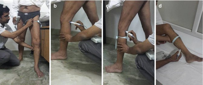
Fig. 6.2
Reflux evaluation by HHD. (a) SF junction. (b, c) SP junction. (d) Medial calf perforator
HHD-Saphenopopliteal Junction
The patient is in the upright position. The limb to be tested is relaxed and kept a little forward with the knee slightly flexed. The examiner is comfortably seated behind the patient. The popliteal vein is insonated with a 5 MHz probe. The response to calf compression and release is observed. A bidirectional flow may indicate either SPJ incompetence or reflux in the popliteal vein. The test is repeated after applying a tourniquet below the probe. If the bidirectional flow is abolished or is reduced significantly, it indicates SP reflux. If it persists even after tourniquet, it is due to reflux in the popliteal vein. HHD evaluation of SPJ is not as sensitive as SFJ (Fig. 6.2b, c).
HHD-Incompetent Medial Calf Perforators
The technique of confirming perforator incompetence using continuous wave Doppler was initially documented by Folse and Alexander. The evaluation of incompetent perforators using HHD is not very sensitive. A competent perforator allows blood to flow from superficial to deep veins during relaxation of calf muscle pump (diastolic phase.). The volume of inward flow of blood may not be sufficient enough to generate a signal. The competent perforator does not permit an outward flow of blood from deep to superficial veins during contraction (systole) of the muscle pump. If this happens, it indicates a dilated and incompetent perforator. Hence, the HHD finding in the presence of an incompetent perforator would be an abnormal signal on calf muscle compression or sometimes a bidirectional flow, if the inward flow is of large volume.
The test can be carried out with the patient in the supine or erect position. We prefer the supine position. The patient lies flat on an examination couch with the knee on the side to be tested flexed at 45–60° and the foot flat on the bed. The other knee is kept straight. A tourniquet is applied over the middle of the leg to prevent any reflux through an incompetent saphenous system. Generous quantity of aqueous gel is applied over a large area on the medial aspect of the leg. A probe of 8 MHz is insonated first over the medial aspect starting just below the medial malleolus and shifting progressively upward along a line 2 cm behind and parallel to the posterior border of tibia (the line of the medial calf perforators). Concurrently, the calf muscle above the tourniquet is compressed and released. The sites of incompetent perforator are indicated at the points where a prominent signal is heard on compression of the calf muscle [11] (Fig. 6.2d).
The deep venous system can be evaluated for patency and reflux using HHD, but the results are not sensitive. Duplex scan would be more appropriate for this purpose.
The limitations of HHD are that the results are observer dependent and are only qualitative. Clinical examination combined with HHD can miss up to 30 % of important connections between deep and superficial veins [2].
Primary Varicose Veins: Investigations
The diagnostic studies used for primary varicose veins broadly falls under two groups: noninvasive studies and invasive studies.
Noninvasive Studies
Duplex Scan
The test of choice for the evaluation of chronic venous disease is duplex scan. This employs a pulse wave Doppler in which the energy of the returning echoes is displayed as an assigned color; by convention, echoes representing flow toward the transducer are seen as shades of red, and those representing flow away from the transducer are seen as shades of blue. The color display is superimposed on the B-mode image, thus allowing simultaneous visualization of anatomy and flow dynamics. It is safe, noninvasive, and cost-effective. The evaluation is objective and quantifiable unlike HHD. It can be repeated for follow-up. Portable units are available for bedside evaluation. It provides simultaneous anatomic and hemodynamic information.
A linear transducer of 5–10 MHz range is ideal for the evaluation of most veins. Higher-frequency probes are used for superficial veins, and lower-frequency probes for evaluation of vena cava and iliac veins. All venous duplex examination includes four components – visualization, compressibility, flow, and augmentation. Duplex ultrasound can be used to demonstrate obstruction and/or reflux. Color coding enables rapid evaluation (Fig. 6.3).
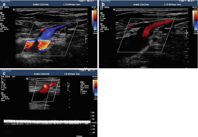
Fig. 6.3
Duplex scan demonstrating gross SFJ reflux. (a) Normal flow, no provocation maneuver, (b) Reflux following provocation. (c) Demonstrating duration of reflux
Duplex scan evaluation of a primary varicose vein patient has the following objectives [2]:
1.
To assess the competence of the SF/SP junctions
2.
The extent and severity of reflux in the axial saphenous veins and also to determine the dimensions of the vein
3.
The site and location of the incompetent perforators
4.
Any nonsaphenous source of reflux
5.
Patency, competence, and any other structural changes in the deep veins
Reflux is the dominant finding in primary varicose veins. It can be elicited by different methods [12].
1.
Response to Valsalva; useful for groin veins
2.


Compression and release distal to point of examination
Stay updated, free articles. Join our Telegram channel
Full access? Get Clinical Tree
