One of the principal goals of preoperative evaluation is to balance the benefit of a curative-intent surgical resection and the risks of immediate and long-term postoperative complications.
While increased perioperative mortality occurs with age >70 (4 vs. 1.4 %, OR 3.6 [95 % CI, 1.4–8.9]) [1], age alone should not be the only determinant of a patient’s operability.
Physiological Consequences of Thoracic Surgery
General to All Surgery
Inhaled volatile agents cause changes in diaphragm and chest wall function, creating areas of decreased ventilation, which leads to ventilation to perfusion (V/Q) mismatch and subsequent hypoxemia.
Specific to Thoracic Surgery
Postoperative chest wall pain causes:
Decreased functional residual capacity (FRC) up to 30–35 % with subsequent atelectasis.
Poor cough with inability to clear pulmonary secretion and increased risk of pneumonia.
Thoracic patients should be evaluated preoperatively to screen for patients at high risk of perioperative complications (Fig. 1.1).
Pulmonary Resection
Impaired FEV1.
Impaired cough.
Atelectasis.
Single-Lung Ventilation
Initially causes 50 % right to left shunt, V/Q mismatch, and hypoxemia [3].
However, compensatory mechanisms in the atelectatic lung decrease perfusion (hypoxic pulmonary vasoconstriction, manipulation of the lung during surgery, and gravitational force from lateral decubitus position). Shunt fraction therefore decreases to 25 %.
Can lead to acute lung injury (ALI) or acute respiratory distress syndrome (ARDS). Risk factors include high tidal volume and airway pressure.
Protective ventilation strategies can decrease injury: low tidal volume ventilation, use of positive end-expiratory pressure (PEEP), and minimizing airway pressure and fraction of inspired oxygen.
Bronchial Anastomoses
Impaired mucociliary clearance and buildup of secretions.
Proximal Foregut Anastomoses
Impaired swallowing and risk of aspiration.
Mitigation of Cardiopulmonary Adverse Events
Cigarette smoking: all active smokers should be encouraged to stop at least 2 weeks before surgery (preferably >6 weeks). Patients should be offered counselling, smoking cessation programs, and pharmacologic assistance.
Estimation of postoperative predicted pulmonary function to assess and stratify risk of pulmonary complications, perioperative morbidity and mortality, and long-term functional disability (Fig. 1.1).
Identification of cardiac patients in need of medical management or coronary revascularization (Fig. 1.2).
Preoperative exercise program in all patients.
Cardiac Assessment
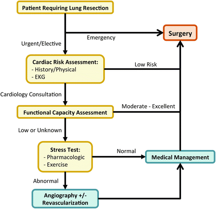
All noncardiac thoracic surgical patients should undergo a cardiac risk assessment based on history, physical examination and baseline electrocardiogram (EKG), and managed according to the 2014 American College of Cardiology/American Heart Association guidelines (Fig. 1.2) [4].
Emergency surgery: proceed to surgery.
Urgent/elective surgery:
If patient is suffering from an acute coronary syndrome, they should be managed according to NSTEMI or STEMI clinical practice guidelines and referred to cardiology.
Evaluate for risk factors of coronary artery disease (CAD) and overall risk of experiencing major adverse cardiac event using risk calculators such as the American College of Surgeons-NSQIP or Revised Cardiac Risk Index. No further testing is needed for patients with low risk (<1 %).
Patients at risk of major adverse cardiac event should be evaluated for functional capacity using an objective scale. No further testing is needed for patients with moderate to excellent functional capacity.
Patients with poor or unknown functional capacity should undergo either pharmacologic or exercise stress testing. Patients with normal stress test can me managed medically and undergo surgery. Patients with abnormal stress test should undergo coronary angiography ± revascularization followed by appropriate medical management prior to proceeding with surgery. Alternatively, a noninvasive surgical approach should be considered (e.g., chemoradiation for malignancy).
Cardiac testing [4]:
Electrocardiogram: indicated in patients with CAD, significant arrhythmia, peripheral arterial disease, cerebrovascular disease or other structural heart diseases. Routine 12-lead EKG is not necessary for asymptomatic patients undergoing low-risk surgical procedures.
Echocardiogram: indicated to rule out asymptomatic pulmonary hypertension in patients who may require pneumonectomy, or in patients with suspected impaired cardiac function, heart failure (e.g., dyspnea of unknown origin), CAD or valvular disease. Patients with previous left ventricular (LV) dysfunction, should also be reevaluated if >1 year since the last evaluation.
Stress testing: indicated in patients with or at high risk for ischemic heart disease in order to identify patients who may require further medical therapy or coronary revascularization prior to thoracic surgery.
Angiogram: indicated to rule out life-threatening coronary stenosis in patients with significantly positive stress tests.
Pulmonary Function Tests (PFT)
Indications:
Significant baseline airflow obstruction with known obstructive pulmonary disease
Significant pleural disease
Central lung mass or suspected endobronchial obstruction
Prior lung resection
Not indicated for surgeries without lung resection (e.g., esophagectomy, mediastinoscopy, pleural biopsy or drainage), and in patients without prior history of lung disease, unexplained dyspnea, or functional limitation.
Baseline Forced Expiratory Volume in 1 Second (FEV1) and Diffusing Capacity of the Lung for Carbon Monoxide (DLCO)
Most commonly used predictors of postoperative pulmonary reserve.
Neoadjuvant Chemotherapy: decreases preoperative and increases perioperative DLCO [5]. Patients who undergo neoadjuvant therapy should undergo repeat PFT after the completion of therapy [2].
Anemia most common cause of falsely low DLCO. Other factors interfering with the interpretation or accuracy of pulmonary function tests include use of bronchodilators, narcotics, pregnancy, increased intra-abdominal pressure (e.g., gastric distension), fatigue, and other conditions limiting the patient’s ability to perform spirometry.
Mortality <5 % with preoperative FEV1 >1.5 L for lobectomy and >2.0 L or >80 % for pneumonectomy [6].
Most patients with FEV1 >60 % will tolerate a lobectomy (morbidity rate 12 %), depending on age and comorbidities [7].
FEV1 is an independent predictor of pulmonary complications and cardiac complications (OR increases by 1.1 for every 10 % decrease in FEV1) [8].
DLCO <70 % has been identified as a predictor of mortality and pulmonary complications [9] and may require additional physiologic testing prior to resection (e.g., exercise testing—VO2 Max).
Stay updated, free articles. Join our Telegram channel

Full access? Get Clinical Tree


