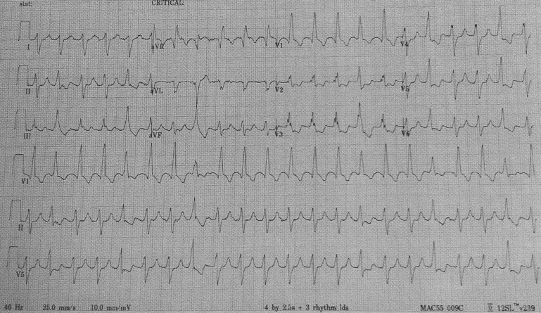
Before cataract surgery, a 64-year-old diabetic man had an electrocardiogram ( Figure 1 ). It shows 257 atrial flutter waves per minute and 2:1 atrioventricular conduction. The second, fifth, eighth, fifteenth, eighteenth, and twenty-first QRSs are fusion complexes of premature ventricular depolarizations with impulses being conducted from the atria. The QRS duration is prolonged (0.13 seconds) with large terminal R waves in leads aVR and V 1 and large terminal S waves in leads I and V 4 to V 6 . These findings indicate complete right bundle branch block, and right axis deviation of the QRS (+141° in the frontal plane) is present as well. An echocardiogram recorded 3 months earlier showed left ventricular dilation with an ejection fraction of 25% and severe inferior wall hypokinesis.