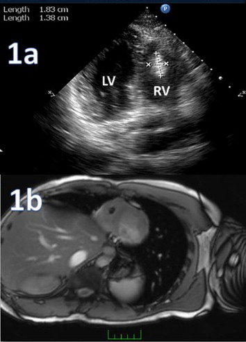
20 years old male patient admitted to our cardiology clinic with stabbing chest pain at a localized point for severeal years. He was just recruited to army and was offered us by his commander for evaluating his physical condition, if he could perform the military exercises. He was not a smoker. His history revealed any disease or specific medical condition. Physical examination revealed ABP:130/85mmhg, pulse 89/min, his heart sounds were rhytmic and there were no additional sound or murmur heard. His ECG revealed sinus rhytm and incompleted riht bundle branch block. His echocardiographic examination revealed nothing abnormal but an 18X13mm right ventricular apical oval hiperecogen mass with smooth borders(1a). Because of the suspected thrombus he was sought for bleeding and coagulation disorders, then for the drug abuse and lastly the constitutional symptoms of any malignity or systemic disease, but not a special finding encountered with. He was referred to cardiac MRI. And contrast enhanced cardiac MRI(1b) reported highly probable thrombus of 9mm diameter localized over the trabecula of right ventricle. Patient then referred to surgical therapy after detailed assesment of ethiological disseases.
Cardiac masses are mostly secondary to other malignities or systemic diseases. Primary ones are the mostly benign cardiac tumours and thrombus,changing according to age and other cardiac or systemic conditions of patients. Transthoracic echocardiography(TTE) is the mostly used and first line diagnostic modality in clinical practice. Becouse of some limitations of variable image quality and artefatcs of TTE there is need for other supportive diagnostic tools for definite diagnose. cMRI is one of the most useful methods for cardiac thrombus. Especially contrast enhanced delayed MRI images are very useful for differentiation of cardiac thrombus from other masses. Because of the avascular structure of thrombus contrast enhanced delayed MRI images show absence of contrast uptake. And for being highly consistent with pathologic reports at the studies, contrast cMRI imaging is an excellent reference technique for differential diagnose of cardiac thrombi from other cardiac masses. Our case is important for reminding the usefulness of contrast enhanced cMRI in the differential diagnose of suspected cardiac thrombus at echocardiography.


