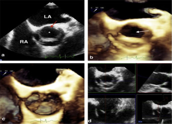
Unicuspid aortic valve(UAV) is a rare congenital cardiac valvular anomaly with an estimated prevalence of 0.02 % in adult patients undergoing transthoracic echocardiographical(TTE) evaluation. It is usually misdiagnosed as bicuspid aortic valve (BAV), which is more prevalent, due to similar symptoms and characteristics during cardiac imaging. Symptoms are related to severe aortic stenosis. As compared to the past when the diagnosis is made during autopsy or intraoperatively, with the advance of cardiac imaging modalities, the diagnosis of UAV is usually established during transthoracic and transesophageal echocardiographic (TEE) evaluation. 26 years old male patient referred to our hospital with the diagnosis of moderate aortic stenosis. Two-dimensional(2D) TTE and TEE evaluation in another hospital reported tricuspid aortic valve with increased aortic velocities (50 and 31 mmHg max and mean respectively). He was reevaluated in our facility. Aortic valve was not consistent with typical bicuspid morphology during 2D TTE and TEE (figure 1a) evaluation; therefore, further evaluation was performed with three-dimensional(3D) TEE (figure 1b and 1c) imaging modalities using multiplane reconstruction (figure 1d) which revealed a characteristic, teardrop-shaped eccentric valve orifice located posteriorly. Consequently the diagnosis of unicommisural unicuspid aortic valve was established with aortic flow velocities consistent with moderate aortic stenosis and regurgitation. The patient was planned to be followed up regularly in outpatient clinic.