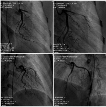
A 32 year-old female was admitted to our hospital for resting squeezing retrosternal chest pain of last month duration and increased last 12 hour’s, and nausea and vomiting. She did not have any coronary artery diseases(CAD) risk factors of excluding smoking and history of known CAD. The patient was hospitalized with a diagnosis of subacute anterior myocardial infarction. On physical examination, arterial blood pressure 107/75 mmHg, pulse 80/bpm, heart and breath sounds was normal. Electrocardiogram (ECG) at admission showed Q wave and minimal ST segment elevation in leads V1-4. Transthoracic echocardiography showed akinesia in the mid-basal segments of anterior left ventricular (LV) wall and hypokinesia in apical segment of LV and ejection fraction %40. Afterwards, the patient underwent coronary angiography (CAG) for the evaluation of coronary arteries. Coronary angiography revealed totally occlusion in proximal segments of left anterior descending artery (LAD) and circumflex (Cfx) and right coronary artery (RCA) normal (Figure 1). The lesion was passed with 0,014 inc floppy wire and performed the balloon angioplasty(3,0x15mm) and TIMI 1-2 flow could be achieved. Subsequently, we performed Intravascular ultrasound (IVUS) examination to investigate the extent of plaque burden. IVUS examination demonstrated minimal plaque burden which no significant stenosis. Unfractionated heparin and tirofiban was started due to diffuse thrombosis raging from the proximal to the distal part of the LAD. The day after, the patient underwent control CAG which revealed that LAD was proximal dissection and narrowed subtotally with TIMI 0-1 flow (Figue 3). Subsequently, we performed IVUS examination which revealed dissection with an intramural hematoma in proximal segment of LAD. TIMI-3 flow distally was achieved after the two stent implantation in dissected segment of LAD(Figure 4). Coronary artery dissection is an important condition which can cause serious problems when cannot recognized and treated in percutaneous coronary intervention. We detected the coronary dissection was developed 24 hour’s after the first intervention and be transformed into intramural hematoma due to antiagregan and anticoagulan theraphy in this patient. The use of IVUS in dessected segment is very effective method in planning of theraphy and stenting in such initiatives.


