(1)
Department of Neuroscience, University of Turin Ospedale Molinette, Turin, Italy
Abstract
Embryologically, the posterior cerebral artery (PCA) is a branch of the internal carotid artery arising from its posterior cranial division; the connection with the basilar artery (pars basilaris) develops later. The connection with the ICA (pars carotica) can completely regress or persist as a large or small vessel, becoming the posterior communicating artery (PcomA). The posterior shift of the vascular territories (posterior temporal and occipital areas) leads to increased distance between the origin of the PCA and its vascular territories. The decrease of this distance by shifting the origin of PCA from the ICA to the BA could be interpreted as a natural evolution to compensate this process (Kier 1974). Variants in this evolution will be described later.
Embryologically, the posterior cerebral artery (PCA) is a branch of the internal carotid artery arising from its posterior cranial division; the connection with the basilar artery (pars basilaris) develops later. The connection with the ICA (pars carotica) can completely regress or persist as a large or small vessel, becoming the posterior communicating artery (PcomA). The posterior shift of the vascular territories (posterior temporal and occipital areas) leads to increased distance between the origin of the PCA and its vascular territories. The decrease of this distance by shifting the origin of PCA from the ICA to the BA could be interpreted as a natural evolution to compensate this process (Kier 1974). Variants in this evolution will be described later.
As it arises from the basilar artery, the PCA can be divided into four segments (Huber 1979) (Figs. 7.1 and 7.4):
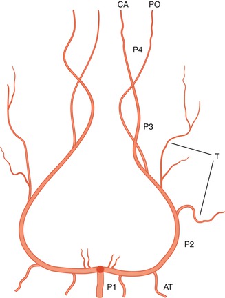
Fig. 7.1
Course of the P1, P2, P3, and P4 segments of the posterior cerebral artery (PCA) in the AP view. Anterior temporal artery (AT), middle temporal and temporo-occipital branches (T), calcarine artery (CA), and parieto-occipital artery (PA)
P1 (precommunicating segment): from its origin from the basilar artery to the junction with the PcomA
P2 (ambient segment): around the midbrain toward the quadrigeminal plate
P3 (quadrigeminal segment): on the surface of the quadrigeminal plate
P4 (distal segments)
7.1 P1 Segment
This is the first segment, extending from its origin in the basilar artery to its junction with the PcomA (Figs. 7.1, 7.2, and 7.4). The P1 segment is the pars basilaris of the PCA that embryologically appears later. It is a short segment, running in the interpeduncular fossa, where it has a close relationship with cranial nerve III, located inferiorly. The P1 segment can have a horizontal course or form a slightly ascending or descending course. Its length varies, with an average of 7.1 mm (Zeal and Rhoton 1978).
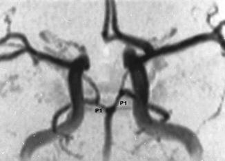
Fig. 7.2
Angio-MRI. The P1 segments are well defined owing to the presence of the posterior communicating artery (PcomA) bilaterally
The P1 segment gives off perforating branches called the thalamoperforating pedicles (Foix and Hillemand 1925a, b) or paramedian thalamic arteries (Percheron 1976a, b). It would be simpler to call these arteries the posterior thalamoperforating branches. They enter the posterior perforated substance (PPS), supplying the medial part of the mesencephalon, the medial part of the thalamus, and the posterior part of the hypothalamus (Lazorthes and Salamon 1971; Saeki and Rhoton 1977; Zeal and Rhoton 1978; Duvernoy 1999; Tatu et al. 2001).
They can also take over the supply of the anterior part of the thalamus, normally supplied by the PcomA, when this is absent (Percheron 1976a, b). On the contrary when P1 is absent, its vascular territory can be supplied by the PcomA or by branches arising from the contralateral P1.
The posterior thalamoperforating branches commonly consist of several small branches, although frequently a larger branch, further dividing into small branches, may predominate. They can arise entirely only from one P1, as previously described (Westberg 1966; Percheron 1976a, b; Saeki and Rhoton 1977; Zeal and Rhoton 1978; Castaigne et al. 1981); this has also been documented more recently (Brassier et al. 1998; Lazzaro et al. 2010). Finally, even with hypoplasia of the P1, perforators may arise from it (Zeal and Rhoton 1978). All these variants probably explain the different pattern of midbrain and thalamic infarction in cases of proximal occlusion of the P1. Examples are presented in Figs. 7.9, 6.5, and 6.11.
The collicular (quadrigeminal) artery, which arises from the P1 and sometimes from the P2 and runs around the midbrain, supplies the lateral and posterior portions of the midbrain. There is frequently a second small artery, running close and parallel to the collicular artery, called the accessory collicular artery (Zeal and Rhoton 1978; Duvernoy 1999; Tatu et al. 2001).
From the P1 and sometimes from the P2 or the parieto-occipital branch arises the posterior medial choroidal artery, which, after reaching the quadrigeminal plate and giving off branches for the posterior thalamus, courses forward in the roof of the third ventricle together with the internal cerebral vein toward the foramen of Monro. It supplies the choroid plexus of the third ventricle (Galloway and Greitz 1960; Wackenheim and Braun 1970; Margolis et al. 1974; Fujii et al. 1980) (Figs. 7.3 and 7.4).
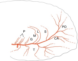
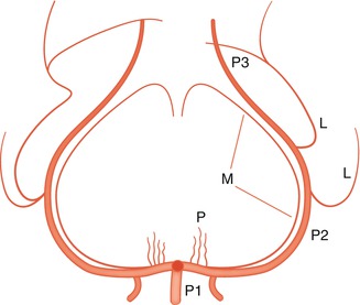
Fig. 7.3
Course of the PCA and its branches in the lateral view. Anterior from PcomA and posterior from P1 thalamoperforating arteries (p), thalamogeniculate artery (G), posterior choroidal arteries, medial (M), lateral (L), splenial artery (S), parieto-occipital arteries (PO), calcarine artery (CA), temporal arteries (T), anastomoses with the anterior cerebral artery (ACA) and middle cerebral artery (MCA) (………)
Fig. 7.4
Course of the P1, P2, and P3 segments of the PCA in the AP view. Posterior thalamoperforating branches (P), medial (M), and lateral (L) posterior choroidal arteries
Near the origin of the P1 arises a meningeal branch. This runs around the midbrain close to and inferiorly to the PCA. The meningeal branch extends to the junction of the tentorium with the falx, vascularizing these dural structures. The meningeal branch is not rare, but it is very fine and so not recognizable on the normal angiogram (Wollschlaeger and Wollschlaeger 1965). It can be involved in pathological processes, especially in the dural arteriovenous fistula of the tentorium (Weinstein et al. 1974) (Fig. 13.14).
7.2 P2 Segment
Also called the circumpeduncular segment, the P2 segment runs around the midbrain (Figs. 7.1 and 7.4), commonly separated from it by several millimeters, and terminates on the surface of the quadrigeminal plate. The P2 segment lies inferior to the optic tract and basal vein, and it is superior to the superior cerebellar artery. The parahippocampal gyrus lies laterally, with the free margin of the tentorium above. The P2 segment gives off the thalamogeniculate artery, which can be a single branch. Sometimes, there are two or three branches, which supply the lateral thalamus (Duvernoy 1999; Tatu et al. 2001). The artery can arise in the proximal or more distal P2 segment (Zeal and Rhoton 1978).
Other branches are the peduncular perforators for the lateral midbrain and the posterior lateral choroidal artery. The latter, which can sometimes arise from the parieto-occipital artery, consists of a single or several branches (Zeal and Rhoton 1978). Basically, there is an anterior branch, which runs in the circumpeduncular cistern, enters the choroid fissure, extends forward to supply the plexus of the temporal horn, and anastomoses with the anterior choroidal artery (AchA). A posterior branch reaches the pulvinar, gives branches to it, and terminates in the choroid plexus of the lateral ventricle at the level of the atrium, anastomosing with branches of the AchA (Galloway and Greitz 1960; Wackenheim and Braun 1970; Margolis et al. 1974; Fujii et al. 1980; Duvernoy 1999) (Figs. 7.3 and 7.4).
From the P2 arise cortical branches for the temporal lobe. They are also called the inferior temporal arteries and supply entirely the inferior surface of the temporal lobe, including the hippocampus and partially the inferior surface of the occipital lobe. The cortical branches for the temporal lobe are classified in relation to the site of origin and vascular territories involved: anterior, middle, and posterior (Zeal and Rhoton 1978). From the posterior temporal artery can arise a few branches supplying the primary visual cortex (Margolis et al. 1974) (Figs. 7.1 and 7.3).
Stay updated, free articles. Join our Telegram channel

Full access? Get Clinical Tree


