20 Pneumothorax

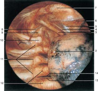
Fig. 20.1
Primary spontaneous pneumothorax. After introduction of the thoracoscope into the sixth intercostal space in the mid-axillary line, the lung is retracted. Visceral and parietal pleura are normal. The view toward the apex of the thoracic cavity shows the upper lobe (1), which appears pinkish at its apex and is air-containing, at its base bluish and atelectatic; between these is a pigmented area and a whitish spot (2) which represents a small ruptured bleb. Subclavian artery (3), phrenic nerve (4), subclavian vein (5), internal thoracic artery (6), first rib (7), superior intercostal artery (8), stellate ganglion at the head of the first rib (9), head of the second rib (10), sympathetic trunk (11), anterior longitudinal ligament (12), intercostal artery (13), intercostal vein (14).

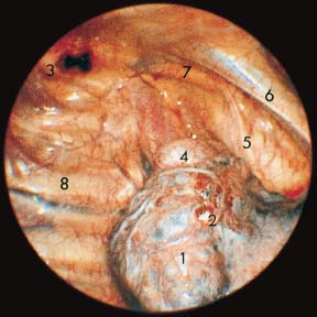
Fig. 20.2
Primary spontaneous pneumothorax. The right upper lobe (1) is bluish and atelectatic, at its apex is an egg-sized bleb (4) and a smaller whitish hemorrhagic strand (2) that is one end of the torn adhesion. At the thoracic apex on the first rib (3), a blackish hemorrhagic area represents the point on the parietal pleura at which the adhesion was attached. The lung has meanwhile separated from the apex of the thoracic cavity. Phrenic nerve (5), subclavian vein (6), subclavian artery (7), chest wall with ribs and intercostal vessels (8).

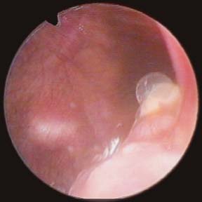
Fig. 20.3
Primary spontaneous pneumothorax. The photograph shows a small apical bleb in a patient with recurrences of pneumothorax. The bleb is not deflated, hence not leaking.

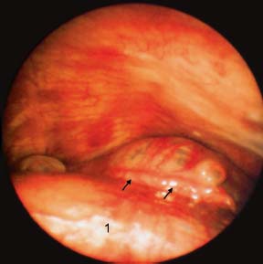
Fig. 20.4
Primary spontaneous pneumothorax. Small bleb (→) at the apex of the left upper lobe (1).
Stay updated, free articles. Join our Telegram channel

Full access? Get Clinical Tree


