15 Pleural Effusions due to Diffuse Malignant Mesothelioma and Asbestos-related Pleural Diseases
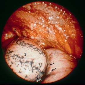
Fig. 15.1
Diffuse malignant mesothelioma (biphasic). After drainage of 3000 mL of hemorrhagic effusion, view toward the apex of the left thoracic cavity. Patchy nodular tumor growth on the left anterior chest-wall pleura (1), larger nodule in the apex (2). Normal-appearing upper lobe (3) with some anthracotic pigmentation and a few tumor nodules at the base (→), small tumor nodules also on the lower lobe close to the fissure ( ).
).
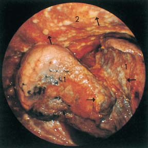
Fig. 15.2
Diffuse malignant mesothelioma (biphasic). After drainage of 3700 mL of hemorrhagic effusion, view toward the apex of the right thoracic cavity. The lung (1) is retracted and compressed; air-containing parenchyma is seen in only a few places. On the visceral pleura (1), the parietal pleura (2), and the diaphragm are numerous white nodules (→).
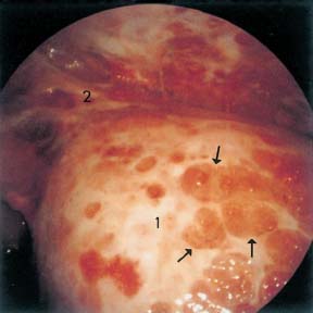
Fig. 15.3
Diffuse malignant mesothelioma (undifferentiated). After drainage of 1500 mL of hemorrhagic effusion, the lung is retracted and covered by a thick pleural peel. The left upper lobe (1) is infiltrated by numerous disklike hyperemic nodules (→) as well as thick adhesions to the chest wall (2), which also contains small hemorrhagic nodules.
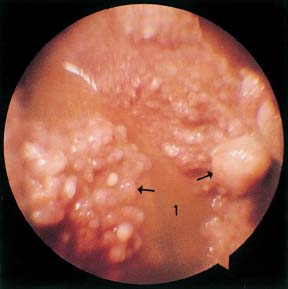
Fig. 15.4
Diffuse malignant mesothelioma (epithelioid). After drainage of 350 mL of gelatinous fluid (1), both pleural surfaces were covered with coarse, irregular grapelike nodules, here on the chest-wall pleura (→).
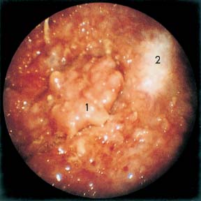
Fig. 15.5
Diffuse malignant mesothelioma (biphasic) with pleural plaque. After drainage of 2000 mL of dark serous fluid, the entire parietal pleura was completely covered by a reddish, irregular layer of tumor from which protruded regular, cauliflower-like whitish tumor nodules (1). In addition, an irregular pleural plaque is seen (2).
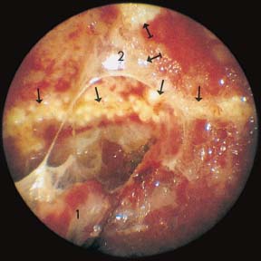
Fig. 15.6
Diffuse malignant mesothelioma (biphasic) with bilateral pleural plaques. After drainage of 1350 mL of hemorrhagic effusion: adhesions between the right upper lobe (1) and the chest wall (2), along the ribs coarse hyaline plaques (→) and, nearby, distinct tumor nodules ( ).
).
Stay updated, free articles. Join our Telegram channel

Full access? Get Clinical Tree


