Fig. 7.1
Chest radiograph showing a trapped and partly deflated left lung following aspiration of pleural fluid with a small bore chest drain. Small right-sided pleural effusion
Video-Assisted Pleural Biopsy and Pleurodesis
Video-assisted thoracoscopy can be carried out under either local anesthesia with sedation or under general anesthesia. It can combine a pleural biopsy, drainage of an effusion and pleurodesis in one procedure. It is not without significant morbidity. It typically requires chest drainage for 3–7 days and a total hospital stay of 7–8 days. About 15 % of patients experience complications such as persisting air leakage, persisting pain, or less commonly pleural infection or pneumonitis [5]. The patient’s overall state of health and the likely benefits need to be considered prior to the procedure.
Indwelling Catheters
Tunneled indwelling pleural catheters are a useful option in those with “trapped lung,” or failed chemical pleurodesis. The catheter can be placed as a day case procedure rather than requiring an inpatient stay in hospital, and this can be of considerable benefit to those with a limited life expectancy. The catheter can be connected to a collecting bottle which is drained as necessary [1]. Complete control of breathlessness is reported in two-thirds of patients. Complications such as loculation of the pleural fluid occur in about 18 % of patients, and infection in about 6 %. Occasionally tumor will seed along the catheter track.
Malignant Mesothelioma
Malignant mesothelioma is a primary tumor of the pleura and occasionally of the peritoneum or pericardium. It is rapidly progressive, surgically incurable, and poorly responsive to radiotherapy and chemotherapy. It affects an increasingly aged population who often have substantial comorbidities. Palliative care is an important consideration for all patients from the time their tumor presents. Pain and breathlessness are the usual presenting symptoms and sweating is often a prominent feature. Psychological and emotional problems are common at the time of diagnosis and as the disease progresses, and patients and their families require substantial support throughout the course of the disease. There are often legal and compensation issues to contend with on account of the tumor’s close association with previous occupational asbestos exposures.
Epidemiology and Pathology
Mesotheliomas are closely associated with work with asbestos and are around 1,000 times more common in those with typical occupational exposures. Asbestos was widely used in transport, construction, and industry in the second half of the twentieth century on account of its structural and insulating properties. The current worldwide mesothelioma epidemic reflects exposures from those sources. Mesotheliomas can occur after radiotherapy and occasionally appear to arise spontaneously but many if not all of these are caused by environmental asbestos exposure.
The risk of mesothelioma increases with the extent of previous asbestos exposure but this is often difficult to quantify accurately and the relationship appears relatively weak. The tumor can occur following low-level exposures, for example, from washing the clothing of a family member. Crocidolite (blue) and amosite (brown) asbestos persist longer in the lung and are considerably more potent causes of mesothelioma than white (chrysotile) asbestos. The relatively low risk associated with the latter may be entirely due to its contamination with another asbestos fiber, tremolite. Industrial workers generally had mixed exposures and the epidemiology of mesothelioma is dominated by the time from first exposure. The risk increases exponentially with time and so mesotheliomas are rare within 20 years of first exposure and occur on average after about 40 years. Most patients are over 70 years of age at presentation. Men are affected five times as often as women reflecting their greater occupational exposures.
Because asbestos exposures came under increasingly stringent control from the early 1970s onwards mesothelioma incidence is currently around its peak in most industrialized countries. In the UK, there are approximately 2,500 cases per annum. The UK rate is five times higher than that in the USA and the total numbers of cases are similar in the two countries despite their different populations. The rates in other European countries are also lower than in the UK, the latter’s high rate being attributed to its greater and more prolonged use of amosite asbestos. Incidences in younger age groups have fallen most in recent years, reflecting better control of their asbestos exposures. As incidence falls, so the average age of those with mesotheliomas will increase and by 2030 most patients are likely to be more than 80 years old. This will influence the approach to the management of the tumor.
Histologically, mesotheliomas can have an epitheliod, sarcomatoid, or a mixed appearance with epithelioid tumors having a better prognosis. Usually they can be differentiated from metastatic pleural carcinomas by their light microscopic appearances and immunohistochemical staining pattern but poorly differentiated tumors are sometimes difficult to characterize. Differentiation from benign reactive pleuritis or pleural fibrosis can be difficult and approximately 10 % of pleural biopsies that show benign features only are falsely negative and subsequently turn out to be malignant [6].
Mesotheliomas usually present with large pleural effusions that cause breathlessness and sometimes chest pain. Occasionally there is solid pleural tumor only. They generally spread diffusely throughout the pleural cavity leading to encasement of the lung. Direct spread to adjacent organs and through the chest wall is common, particularly along the tracks of biopsies and through incisions. Distant metastases are frequently found at autopsy but they rarely give rise to symptoms. Progression is usually rapid with median survival in the region of 12 months for epithelioid and 6 months for sarcomatoid tumors [7]. A recent study from Australia confirmed the survival difference between epithelioid and sarcomatoid tumors [8]. It also showed that those with peritoneal mesothelioma had a worse median survival, at 16 weeks, than pleural (36 weeks). Finally, it reported an improvement in survival by decade from 25 weeks in 1970–1979 to 43 weeks from 2000 to 2005. This may be due to diagnosis of the tumor at an earlier stage. A few patients survive for several years and untreated survival of up to 10 years has been reported.
Presentation and Symptoms
Given its doubling time, most mesotheliomas are likely to have been present subclinically for 10 years or so by the time of diagnosis. Occasionally the tumor is identified on a radiograph or CT scan obtained for some other purpose but for most individuals it is the abrupt onset of breathlessness associated with a pleural effusion and chest pain that brings the tumor to light. Abdominal symptoms predominate in peritoneal mesotheliomas.
The presenting symptoms in patients with pleural mesothelioma, are mostly physical. Because of the subsequent realization of its association with previous occupation and the poor prognosis, emotional issues become more prominent with the progression of the disease. These affect both patient and carer.
Presenting Symptoms
In early disease, local symptoms predominate over systemic ones. Of these, pain and breathlessness are the commonest, but cough is also described. The prevalence of these symptoms varies in different series [9]. Pain has been reported in 29–85 %, breathlessness in 28–89 %, and both in 89–96 % of patients at presentation. Cough is reported in 16–75 %. The most prominent systemic symptoms are fatigue, anorexia, weight loss, and sweating. These tend to become more prominent as the disease progresses.
The pain of mesothelioma can be nociceptive, neuropathic, or sympathetically maintained. As the tumor usually originates in the parietal pleura, localized opioid sensitive pain is common, particularly if there is no associated pleural effusion. As it progresses, the tumor invades surrounding structures. Outwardly this involves the chest wall with compression or destruction of the intercostal nerves, resulting in neuropathic pain, as well as muscle, ribs, subcutaneous tissues, and skin. Medially the mediastinal structures and vertebral column can be affected. At the thoracic inlet, involvement of the brachial plexus results in neuropathic pain, and involvement of the subclavian sympathetic chain causes sympathetically maintained pain of the arm.
Breathlessness is usually initially due the presence of a pleural effusion, and responds to removal of that fluid. With disease progression the pleural tumor encases and restricts expansion of the affected lung. When this happens, more general dyspnea-management strategies are needed. Mesothelioma can also affect the pericardium, causing pericardial effusion or tamponade. This can be managed acutely by pericardial aspiration or for the longer term by the formation of a pericardial window. A less common cause of breathlessness is phrenic nerve damage, due to mediastinal invasion, resulting in diaphragmatic paralysis.
Cough is due to pleural irritation, and is usually dry. It is commonly encountered towards the end of pleural aspiration and is often the indicator to halt the procedure. Opioids are the drug treatment of choice for this type of troublesome cough.
Although mesothelioma can and does metastasize via the bloodstream, symptoms of distant spread rarely predominate over local ones. A more likely complication of advancing disease is of penetration of the tumor through the diaphragm with ascites and intra-abdominal lymphadenopathy.
Systemic Symptoms
Emotional symptoms are common. These include anxiety, fear, anger, and depression. The prevalence of these has been reported at between 20 and 67 % in patients and 57 to 84 % of carers [10]. Anorexia and weight loss are less common at presentation, but tend to become more prevalent as the disease progresses. Prevalence has been reported at between 3 and 87 %. The symptoms are probably a manifestation of the anorexia/cachexia syndrome rather than starvation due to loss of appetite with subsequent low calorific intake alone.
Fatigue is another symptom which tends to become more prominent with progressive disease. It has been reported in 32–94 % of patients. Fever and sweats seem to be more prevalent in mesothelioma patients than in those with lung cancer. Fever has been reported in 3–9 % patients at diagnosis and sweats in 20 %. Again the symptoms worsen with advancing disease. Details of the management of these symptoms are given in Chap.6.
Approach to the Patient with Suspected Mesothelioma
The management of malignant mesotheliomas is primarily palliative. They are rapidly progressive, are almost universally fatal, and have no treatment that markedly prolongs life. The typical patient is elderly and has comorbidities. Fewer than 50 % of patients have World Health Organization performance status of 0 or 1 at presentation, indicating that they are unable to carry out the equivalent of light housework [7]. A quarter have comorbidities sufficiently severe to preclude any form of active treatment. An early balance may have to be struck between undertaking invasive investigations and living with diagnostic uncertainty, between symptom control and attempts at modifying the course of the disease, and between maintaining a positive approach and tempering unrealistic expectations.
The diagnosis of mesothelioma may be strongly suspected if someone with a history of asbestos exposure or asbestos pleural plaques on a chest radiograph presents with a unilateral pleural effusion, particularly if that is associated with chest pain and some weight loss. The history of asbestos exposure may need to be actively sought as many patients initially deny exposure and only recall it later after detailed enquiry. The relevant exposures may have been brief, may have occurred indirectly from other workers using asbestos in the vicinity or even from domestic contact, and may have occurred more than half a century earlier. The risk of developing a malignant mesothelioma is in the region of 2 % for most asbestos-exposed workers though historically some groups who were heavily exposed to crocidolite asbestos had much higher rates than that.
Even if the diagnosis of mesothelioma is strongly suspected, alternatives such as cardiac failure, tuberculosis, or rheumatoid pleural disease need to be considered as their management is very different from that of suspected pleural malignancy. More often the principal differential diagnoses will be of a benign asbestos-induced pleural effusion or a secondary pleural adenocarcinoma. A number of very rare primary pleural sarcomas are described.
Asbestos can cause benign pleural effusions that resolve, leaving diffuse pleural fibrosis. The risk for typical asbestos-exposed workers is around 2 %, which is similar to their risk of developing a mesothelioma. Benign asbestos-induced pleural effusions can be asymptomatic but they are often associated with breathlessness. They can be clinically very difficult to distinguish from mesotheliomas, though they are less likely to be associated with chest pain or systemic malaise. They frequently present sequentially, affecting one side and then after an interval of several months the contralateral side. That helps clarify the diagnosis as mesotheliomas are almost never bilateral. There is no effective treatment.
The pleural cavity is a common site for metastases from lung and breast tumors, and occasionally from stomach, kidney, ovary, and prostate cancers. Metastatic pleural adenocarcinomas often present with pleural effusions and appear clinically and radiologically similar to mesotheliomas. The pleural involvement renders the primary tumor surgically incurable though it may still be amenable to chemotherapy. Thus, even if the pleural disease appears clearly malignant, obtaining a histological diagnosis may help guide treatment.
Diagnostic Investigations
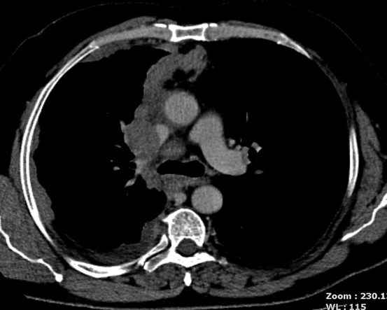
A chest radiograph will almost always be obtained from a patient presenting with a suspected pleural malignancy. A diagnostic pleural aspirate can be easily obtained in the out-patient clinic but the test is relatively insensitive, identifying malignant cells in only 30–50 % of mesotheliomas [11]. Computed tomography (CT) is associated with little additional morbidity and should generally be considered. Features such as nodularity of the pleural shadowing or extension of pleural shadowing onto the mediastinal surface suggest malignancy rather than benign disease though they do not differentiate mesotheliomas from metastatic pleural tumors (Fig.7.2). If there is an identifiable pleural mass, it can be biopsied under CT guidance and this will identify around 80–85 % of mesotheliomas. If breathlessness is a significant problem and the patient’s general physical condition allows it, then a thoracoscopy with pleural aspiration, biopsy, and pleurodesis is likely to be the most rapid way of achieving a diagnosis and symptom control.
 < div class='tao-gold-member'>


Only gold members can continue reading. Log In or Register to continue
Stay updated, free articles. Join our Telegram channel

Full access? Get Clinical Tree


