Pharmacologic Prophylaxis for Post-cardiopulmonary Bypass Bleeding


Pharmacologic Prophylaxis for Post-cardiopulmonary Bypass Bleeding


Niamh A. McAuliffe

Deepak Hanumanthaiah

C. David Mazer


INTRODUCTION

Blood transfusion in cardiac surgery patients has warranted and generated much investigation and discussion. Significant perioperative blood loss requiring transfusion of blood products has been associated with increased morbidity and mortality in cardiac surgery (1). In the quest for reducing the inherent risks and significant costs of transfusion, many aspects of the perioperative course have been examined. Factors which contribute to the requirement for blood transfusion in the perioperative period include preoperative antiplatelet medication, the nature of the surgery, hemolysis by extracorporeal circuits, hemodilution, and heparin use. Up to 50% of patients undergoing cardiac procedures receive no blood transfusion (2). However, there is a subset of “high risk” patients who require substantial perioperative blood transfusion. One review suggested an overall blood transfusion rate of 57% in patients with a packed red blood cell (RBC) transfusion rate of 49% and a non-RBC component transfusion rate of 27% (3). An audit by the European Association for Cardio-thoracic Surgery estimated that cardiac surgery accounted for 5% of blood donated in the United Kingdom (4). Perioperative transfusion of packed RBCs has been associated with increased risk for a broad spectrum of postoperative morbidity after cardiac surgery, including renal failure, prolonged ventilation, infection, neurologic events, and mortality (1). Karkouti et al. (5) demonstrated that massive blood loss after cardiac surgery resulted in an 8-fold increase in mortality. Blood transfusion of even minimal amounts has been associated with increased morbidity and mortality for on-pump coronary artery bypass surgeries (6). Studies have linked perioperative blood transfusion during cardiac surgeries with mediastinitis leading to increased cost and mortality (7). Increased risk of delirium in the postoperative period has been linked to duration of storage of packed RBCs (8). A review of 42 studies showed an association between intraoperative blood transfusion and wound infection in cardiac surgical patients (9). In a large prospective study, Horvath et al. (10) identified pneumonia and bloodstream infections as the most common manifestations of infection and the incremental risk associated with each unit of packed RBCs was 29%. Since the early 1990s, antifibrinolytic drugs have played a major role in blood conservation strategies in cardiac surgery. Perioperative blood conservation strategies play an important role in the reduction of blood transfusion. This chapter will focus on the key prophylactic role played by antifibrinolytic drugs in reducing blood loss and transfusion.


FIBRINOLYSIS AND ANTIFIBRINOLYTIC MECHANISMS

Hemostasis is the process of clot formation at the exposure site of subendothelial collagen in a blood vessel. The hemostatic system consists of four components: coagulation system, endothelium and regulatory proteins, platelets, and fibrinolysis (11). Nonendothelial surfaces in the cardiopulmonary bypass (CPB) circuit initiate contact activation of the intrinsic coagulation pathway resulting in nonhemostatic thrombin generation (12). Under normal hemostatic conditions, soluble fibrin accounts for 1% of total fibrin formation. CPB leads to dysregulation of fibrin resulting in an increase in soluble fibrin of up to 35% (13). Thrombin generation and soluble fibrin formation increase at initiation of CPB, at CPB cessation, and after protamine administration (14). High-dose unfractionated heparin, via antithrombin III, decreases thrombin activity and fibrin generation, thereby preventing clot formation within the extracorporeal circuit (15). However, thrombin has also been widely recognized as a key amplifier protein of inflammation, coagulation, and fibrinolysis. Thrombin causes the release of tissue plasminogen activator (tPA) from endothelium which promotes plasminogen conversion to plasmin, an enzyme that proteolytically degrades fibrin (16). The plasminogen molecule contains specific lysine-binding sites that interact with both fibrin and the major inhibitor, α2-antiplasmin. Urinary plasminogen activator (uPA) and tPA are the two main serine protease activators (17). Vascular endothelial cells secrete tPA, while uPA is secreted by a variety of cell types including nonvascular cells. Bradykinin has been shown to play an important role stimulating the release of tPA via β2 receptors (18). Activators convert plasminogen to plasmin, generating soluble degradation products and exposing carboxy-terminal lysine residues (19).

Other factors contribute to the initiation of fibrinolytic activity during cardiac surgery. Surgical trauma, such as sternotomy alone, can activate fibrinolysis. There is contact activation of fibrinolysis by the CPB circuit, resulting in a 5-fold increase
in tPA activity (14). Peak tPA levels occur early during CPB within 30 to 60 minutes. Increased soluble fibrin is a powerful stimulator of tPA (12). Unfractionated heparin in itself is also a mild stimulator of vascular plasminogen activators, thereby contributing to fibrinolytic augmentation (20). This hyperfibrinolytic state consumes fibrinogen, thereby impairing coagulation postoperatively. In addition to fibrin, plasmin cleaves fibrinogen and a variety of plasma proteins. The breakdown of fibrin strands by plasmin results in the release of fibrin degradation products that include D-dimers. D-Dimer levels have been used as indicators of fibrinolysis in many studies.


FIGURE 21.1. Site of action of antifibrinolytics. Fibrin is polymerized by thrombin and activated Factor XIII at the site of vascular injury. Thrombin-activated fibrinolysis inhibitor (TAFI) helps stabilize the clot against the activity of plasmin. Broken lines indicate inhibitory action of the protease inhibitors. (Reused from Ide M, Bolliger D, Taketomi T, Tanaka KA. Lessons from the aprotonin saga: Current perspective on antifibrinolytic therapy in cardiac surgery. J Anesth 2010;24(1):96-106).α-2 AP-α-2 antiplasmin; tPA- tissue plasminogen activator; TAFIa- activated TAFI; Plgn-plasminogen; PC- Protein C; APC- Activated Protein C; AT- Antithrombin; PAR- Protease-activated Receptor1.

Plasmin activity is regulated both by plasminogen activators and inhibitors. Inhibitors of plasminogen activation are plasminogen activator inhibitor-1 (PAI-1), α2-antiplasmin, and thrombin-activated fibrinolysis inhibitor (TAFI). PAI-1 is a member of the serine protease inhibitor family. It is produced by vascular endothelial and smooth muscle cells and is the primary physiologic regulator of uPA and tPA activity (21). PAI-1 is an acute phase reactant and levels are increased following surgery (14). α2-antiplasmin is manufactured in the liver and is the major inhibitor of plasmin in plasma. TAFI is activated by thrombin and removes carboxy-terminal lysine residues, thereby attenuating plasmin generation. The resultant overall hyperfibrinolytic state consumes fibrinogen impairing coagulation postoperatively, and increasing postoperative hemorrhagic complications and blood transfusion requirements (22). When antifibrinolytic agents are prophylactically administered during CPB, they reduce the susceptibility of fibrin clots to plasmin-mediated degradation.

Plasmin and plasminogen bind at lysine-binding sites onto fibrinogen. The lysine analogs e-aminocaproic acid (ACA) and tranexamic acid (TXA) competitively inhibit the fibrin-binding site on plasminogen and prevent the degradation of fibrin and the dissolution of clot (Fig. 21.1, Table 21.1).


Tranexamic Acid

TXA is an isomer of 4-aminomethylcyclohexane carboxylic acid which competitively inhibits plasminogen. TXA binds to both strong and weak lysine-binding sites on plasminogen and has poor affinity for other plasma proteins. Saturation of the lysine-binding sites of plasminogen with TXA displaces plasminogen from the fibrin surface. At a concentration of 10 µg/mL, 80% of plasminogen was inhibited; with higher doses, 100% of plasminogen activity was inhibited with a plasma concentration of 100 µg/mL (23). At higher concentrations, it is also a noncompetitive inhibitor of plasmin (24). Another proposed mechanism of TXA action is that it increases collagen synthesis, thus increasing the tensile strength of the clot (25). The reduction in D-dimers following TXA use has been used as an indicator of reduced fibrinolysis (26).

The molecular formula of TXA is C8H15NO2. It is a weak acid (pKa ˜4.3), which does not bind to plasma proteins and is nonlipophilic. Administration can be via oral, intravenous, or topical routes (27). Absorption after oral ingestion is approximately 50% and the half-life is approximately 80 minutes (28). A two-compartment model best describes the distribution of TXA (29). The following pharmacokinetic properties for a 70-kg adult were documented by Grassin-Delyle et al. (30): peripheral volume of distribution = 10.8 L, volume of central compartment = 6.6 L, clearance = 4.8 L/hr, diffusional clearance = 32.2 L/hr. Two covariates (body weight and creatinine clearance) are known to affect drug levels. Although CPB was previously thought to affect TXA pharmacokinetics, recent research suggests that modern CPB techniques do not have a significant effect (30,31,32). Up to 95% of TXA is excreted unchanged in the urine, with the remainder metabolized by biotransformation involving acetylation and deamination. As renal excretion of TXA is directly related to creatinine clearance, renal failure may prolong its half-life and
thus potentially lead to increased serum concentrations when TXA is administered by continuous infusion. Various suggestions for reduction in TXA infusion rates in the setting of renal dysfunction are shown in Table 21.2.


TABLE 21.1. Comparison of antifibrinolytic drugs


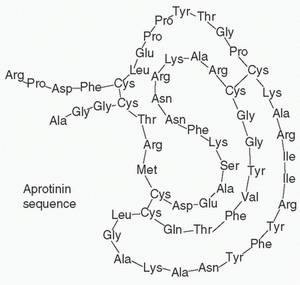
Aprotinin


Aminocaproic acid


Tranexamic acid


Structure


image


image


image


Chemical formula


C284H432N84O79S7


C6H13NO2


C8H15NO2


Molecular weight


6,512


131


157


Plasma level (mg/dL)


4.2


60


3.3


Ki values (Mol.)


Plasmin


7 × 10-11


3.2 × 10⁻¹


1.6 × 10⁻²


Kallikrein


3.6 × 10⁻⁸


NS


NS


Thrombin


6.1 × 10⁻⁵


NS


NS


FXIa


1.1 × 10⁻⁶


NS


NS


APC


1.1 × 10⁻⁶


NS


NS


NS, not significant; Ki, inhibition coefficient; FXIa, activated Factor XI; APC, activated Protein C.


It has been suggested that the optimal benefits from TXA use are derived when administration is initiated at commencement of surgery and maintained throughout by continuous infusion (33). However, others have reported beneficial effects with bolus dosing only. In addition, the timing of drug initiation varies from immediately after induction of anesthesia to after heparin administration. Advocates of the former approach suggest that fibrinolysis begins as early as surgical incision, whereas advocates of the latter approach suggest that heparin may protect against potential thrombotic effects of antifibrinolytic agents. Fiechtner et al. (34) demonstrated that inhibition of fibrinolysis could be achieved with a bolus of 10 mg/kg followed by an infusion of 1 mg/kg/hr, which results in a plasma concentration of 25 to 40 µg/mL, whereas Dowd et al. suggested that higher doses (30 mg/kg bolus, 2 mg/kg added to CPB prime and 16 mg/kg/hr; the “Blood Conservation Using Antifibrinolytics in Randomized Trial [BART] dose”) were required to maintain TXA concentrations greater than 800 µmol/L (29). Many dosing regimens have been studied (Table 21.3), however the optimal dose has not been established. Although some advocate lower TXA dosing regimens to avoid the potential side effects (i.e., seizure activity), two recent randomized, controlled studies have demonstrated significantly reduced bleeding and/or transfusion with the high-dose (BART) regimen (35,36). In both of these studies, there was no significant difference in the incidence of seizures with the BART regimen compared with lower-dose regimens.

Another area of interest is the topical use of antifibrinolytics to reduce postoperative bleeding. Topical use of TXA,
aminocaproic acid, and aprotinin have all been described. For TXA, the dosing regimens have consisted of administration of 1 to 2.5 g in 100 to 250 mL into the pericardial cavity at the time of surgery, either alone or combined with systemic administration. The theoretical advantage of topical administration is targeted local effect with avoidance of any systemic absorption and systemic side effects (37). Some small individual studies have shown a trend toward decreased bleeding and transfusion requirements (38,39,40,41); meta-analysis suggests significant reduction in chest tube losses and transfusion rates (42,43).


TABLE 21.2. Suggested reduction in tranexamic acid maintenance infusion rates in renal disease


Serum creatinine (mg/dL)a


% of usual maintenance dosea


% of normal creatinine clearanceb


% of usual maintenance doseb


1.6-3.3


75


75


75


3.3-6.6


50


50


37.5


>6.6


25


25


31.3


10


10


1


1


a Grassin-Delyle S, Tremey B, Abe E, et al. Population pharmacokinetics of tranexamic acid in adults undergoing cardiac surgery with cardiopulmonary bypass. Br J Anaesth 2013;111:916-924; Fiechtner BK, Nuttall GA, Johnson ME, et al. Plasma tranexamic acid concentrations during cardiopulmonary bypass. Anesth Analg 2001;92:1131-1136.

b Yang Q, Jerath A, Bies RR, et al. Pharmacokinetic modeling of tranexamic acid for patients undergoing cardiac surgery with normal renal function and model simulations for patients with renal impairment. Biopharm Drug Dis 2015. doi:10.1002/bdd.1941.


TABLE 21.3. Reported dosing regimens for tranexamic during cardiac surgery


Only gold members can continue reading. Log In or Register to continue

Stay updated, free articles. Join our Telegram channel

Jun 7, 2016 | Posted by in RESPIRATORY | Comments Off on Pharmacologic Prophylaxis for Post-cardiopulmonary Bypass Bleeding

Full access? Get Clinical Tree

Get Clinical Tree app for offline access

Study name or first author


Journal name and year


Bolus dose


CPB dose


Infusion rate


Serum TXA concentration


BART (81)


N Engl J Med, 2008 Anaesthesia, 2012


30 mg/kg


2 mg/kg


16 mg/kg/hr


>100 µg/mL


Grassin-Delyle (30)


Br J Anaesth, 2013


46 mg/kg


9-11 mg/kg/hr


150 µg/mL


Bokesch (35)


J Thorac Cardiovasc Surg, 2012


1 g


500 mg


400 mg/hr


>600 µmol/L


Fiechtner (34)


Anesth Analg, 2001


10 mg/kg


1 mg/kg/hr


20-70 µg/mL


Karski (112)


J Cardiothorac Vasc Anesth, 1998


50-150 mg/kg


Dietrich (113)


Anesth Analg, 2008


2 g


2 g


1 g/hr


Karski (114)


J Thorac Cardiovasc Surg, 1995


10 g


2 g/hr (for 5 hr)


Horrow (115)


Anesthesiology, 1995


10-40 mg/kg


1-4 mg/kg/hr


Abrishami (42)


Can J Anesth, 2009


1-2.5 g (topical route)