Pericardial Cysts and Cystic Lesions
Allen P. Burke, M.D.
Mesothelial (Pericardial) Cysts
Mesothelial cysts account for ˜20% to 30% of mediastinal cysts. Bronchogenic cysts are more common, but are usually paratracheal and outside the pericardial sac (see below).1
Pericardial cysts are mesothelial-lined structures that are either congenital or acquired and pose no diagnostic difficultly by imaging, unless there are superimposed degenerative or infectious changes. The mean age at presentation is about 50 years. Pericardial cysts may be incidentally discovered on chest x-ray or other imaging or cause symptoms of dyspnea, chest pain, or cough. The typical location is on the right side near the cardiophrenic angle and occasionally the left cardiophrenic angle.1
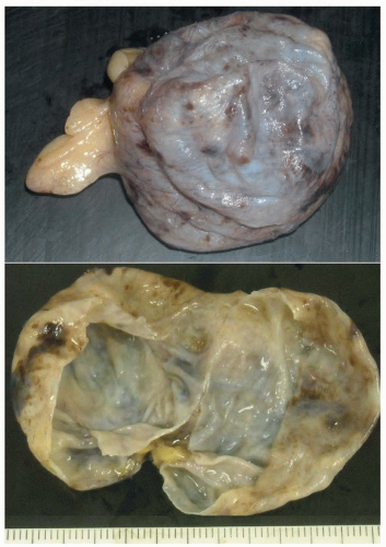 FIGURE 186.1 ▲ Pericardial (mesothelial) cyst. There is a multiloculated thin-walled cyst, which contains serous fluid. The patient was a middle-aged woman with atypical chest pain; CT scan showed a large cystic lesion in the right pericardium. |
Grossly, the specimen will either be in pieces, and demonstrate nondescript fragments of fibrovascular tissue, or be removed intact, in which case there is a simple cyst with serous fluid (Fig. 186.1).
Histologically, the cysts are lined by mesothelial cells (Fig. 186.2), which can be confirmed by staining with anticalretinin, which is usually unnecessary. There may be fibrosis and chronic inflammation, with cholesterol clefts, if there was prior infection or inflammation, which occurs in a minority of cases.
Pericardial Lymphangioma
Also referred to as hygromas, lymphangiomas are composed of dilated lymphatic channels arising from sequestered lymphatic tissue, which fails to communicate normally with the lymphatic tree. Lymphangiomas are uncommon in the mediastinum, representing only 2% of mediastinal cysts.2 Intrapericardial lymphangiomas are extremely rare and occur in infants, children, and young adults. Grossly, the surface is smooth with a bosselated surface. The histologic appearance of intrapericardial lymphangioma is identical to lymphangiomas of other soft tissue sites.
In contrast to mesothelial pericardial cysts, lymphangiomas are multilocular, are lined by flattened endothelial cells, contain proteinaceous fluid and lymphocytes, and often have smooth muscle cells and lymphoid aggregates within the cyst wall (Fig. 186.3).
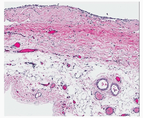 FIGURE 186.2 ▲ Pericardial (mesothelial) cyst. Histologically, the cyst wall resembles normal pericardium.
Stay updated, free articles. Join our Telegram channel
Full access? Get Clinical Tree
 Get Clinical Tree app for offline access

|