Percutaneous Transcatheter Closure of Patent Foramen Ovale


Percutaneous Transcatheter Closure of Patent Foramen Ovale


Ivan P. Casserly

Murat E. Tuzcu


Persistent patency of the foramen ovale has been implicated in an increasing number of pathologic states. Most notably, paradoxical embolism of venous thrombus across the patent foramen ovale (PFO) has been suggested as the leading pathogenic mechanism of ischemic stroke or transient ischemic attacks (TIA) in patients with no other identifiable cause; this is termed “cryptogenic stroke” (1, 2, 3). The potential scope of this clinical problem is underscored by data suggesting that 70,000 cryptogenic strokes are associated with a PFO each year in the United States (4). Percutaneous closure of these defects seems a logical therapeutic strategy. In the late 1980s and early 1990s, the development of closure devices for the nonoperative closure of atrial septal defects quickly found application in the closure of PFOs (5), and additional devices specifically designed for PFO closure subsequently have been developed.

Adult interventional cardiologists have enthusiastically embraced the therapeutic strategy of percutaneous PFO closure. Unfortunately, this enthusiastic acceptance masks a real deficiency of data in a number of key areas: (a) the causal relationship between PFO and cryptogenic stroke remains unproven, and (b) assuming a causal relationship exists, the therapeutic effectiveness of percutaneous closure over current medical therapy in preventing recurrent events is unknown. The result is that appropriate patient selection often presents a greater challenge than the technical aspects of the procedure. The success of a percutaneous PFO closure procedure is founded on the use of good clinical judgment in patient selection, an understanding of the embryology and anatomy of the PFO and atrial septum, an appreciation of echocardiographic findings that impact procedural technique and strategy, and an attention to a number of key fundamental principles during performance of the procedure.


EMBRYOLOGY AND ANATOMY

In utero, the foramen ovale describes the opening between the free margin of the septum secundum and the endocardial cushion (Fig. 24.1) (6). The septum primum lies to the left of the septum secundum and forms the floor of the foramen ovale. Uterine blood is diverted from the inferior vena cava toward the foramen ovale and across the channel that is created by the overlap of the septums primum and secundum. Following birth, the septum primum typically fuses with the septum secundum, thus eliminating the communication between the two chambers. Postmortem and transesophageal echo (TEE) studies have demonstrated that this fusion is incomplete in approximately 25% of individuals, resulting in a PFO (Fig. 24.2). PFOs are identified as a crescentic-shaped tunnel-like opening in the superior portion of the fossa ovalis; this opening has a valve-like property, selectively allowing right-to-left shunting across the atrial septum.

Atrial septal aneurysms (ASA) represent a further anomaly of the atrial septum, and they are caused by an abnormality of the septum primum portion of the septum. The classical appearance of an ASA by echocardiography is that
of a thin hypermobile redundant membrane in the area of the fossa ovalis. In practical terms, a septum with a maximal excursion into either the right and/or left atria of > 10 mm is typically used to define the presence of an ASA. ASAs are much less common than PFOs, with an incidence (as assessed by TEE) of approximately 2%, and an associated PFO is found in >50% of these patients.


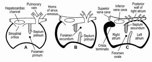
Figure 24.1. Embryologic development of the atrial septum as viewed in cross-section (A-C. See text for details.) (Reproduced with permission from Fitzgerald MJT. Thoracic organs. In: Fitzgerald MJT, ed., Human embryology. Maryland: Harper & Row, 1976:83-105.)


PATIENT SELECTION


Prevention of Paradoxical Embolism

The dominant indication for percutaneous PFO closure is the prevention of recurrent events following an initial presentation with cryptogenic stroke or TIA. Deciding which of these patients warrant percutaneous PFO closure is a significant challenge. Three fundamental questions should be asked before proceeding with this procedure:


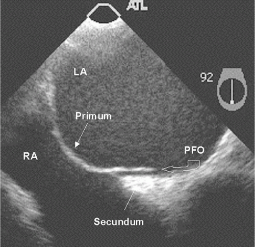
Figure 24.2. TEE images of patent foramen ovale (PFO). This PFO has a tunnel-like appearance. RA, right atrium; LA, left atrium.


1. In this patient, is there reasonable evidence of a causal relationship between the PFO and the cryptogenic stroke or TIA?

The etiology of cryptogenic stroke or TIA is heterogenous, and paradoxical embolism across an associated PFO represents the pathogenic mechanism in only a subgroup of these patients. Therefore, the association of a PFO in patients with cryptogenic stroke or TIA may be incidental or causal. Except in rare circumstances when thrombus is visualized crossing the PFO (7), or a cryptogenic stroke or TIA coincides with the diagnosis of deep venous thrombosis (DVT) (8), the nature of the association in an individual patient is a matter of judgment based on the synthesis of clinical, imaging, and laboratory findings (Table 24.1).


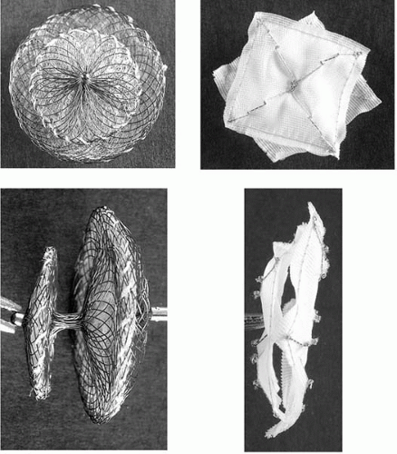
Figure 24.3. Photographs of the Amplatzer PFO occluder (left) and CardioSEAL (right) devices en face and in side profile.


TABLE 24.1. LIST OF CLINICAL, IMAGING, AND LABORATORY VARIABLES THAT FAVOR THE ROLE OF PARADOXICAL EMBOLISM ACROSS A PFO AS THE ETIOLOGY OF CRYPTOGENIC STROKE


Clinical


Age <55 years (2)


Absence of atherosclerotic risk factors, documented atherosclerosis, structural heart disease, or history of atrial fibrillation


Laboratory


Negative hypercoagulable panel


Imaging


CT head


Multiple strokes in multiple cerebral territories


Echocardiography of atrial septum


Large anatomic size of PFO


Large right-to-left shunt at rest or minimal provocation


Associated atrial septal aneurysm


2. What is the likelihood of a recurrent cerebral ischemic event in this patient?

A prospective multicenter French study of 581 young patients (i.e., 18 to 55 years) with cryptogenic stroke treated with aspirin (300 mg/day) provides the best available data on the likelihood of recurrent cerebral ischemic events in cryptogenic stroke patients on standardized medical therapy (9). At 4-year follow-up, the risk of recurrent stroke among patients with no atrial septal abnormality was 4.2% (CI 1.8% to 6.6%), and in patients with PFO alone was 2.3% (CI 0.3% to 4.3%). The combination of PFO and ASA resulted in a dramatic increase in the risk of recurrent stroke at 4 years to 15.2% (CI 1.8% to 28.6%).

These data highlight a number of important points. The occurrence of recurrent events in patients without atrial septal abnormalities emphasizes the importance of non-PFO-related factors in cryptogenic stroke pathogenesis and reinforces the heterogeneity of this diagnosis. In younger cryptogenic stroke patients on medical therapy (either aspirin or warfarin), the presence of a PFO alone is associated with a low rate of recurrent events that is roughly equivalent to that in patients without any atrial septal abnormalities. In contrast, the combination of PFO and ASA appears to dramatically increase the risk of recurrent events. Beyond the presence of an associated ASA, larger anatomic and functional PFOs are assumed to predict an increased risk of recurrent events on the basis of the increased risk of incident cryptogenic stroke in patients with these features. Definitive prospective data supporting the latter assumption are lacking however.


3. What is the optimal therapy in patients with cryptogenic stroke and PFO?

Three strategic approaches to therapy can be taken in patients who present with cryptogenic stroke and are found to have a PFO: medical therapy, percutaneous closure, and surgical closure. With the availability of percutaneous PFO closure, surgical closure is rarely practiced, except when the patient is undergoing open-heart surgery for another primary indication. The clinically relevant question, therefore, is how percutaneous closure compares with medical therapy. To date, no randomized comparison between these treatment strategies has been completed and nonrandomized comparisons are inherently flawed. Unfortunately, this task is complicated by the lack of consensus regarding the optimal medical therapy in patients with cryptogenic stroke. The PICSS substudy showed a trend in favor of warfarin compared with aspirin (10), but the applicability of these results to a more typical younger cryptogenic stroke population is uncertain.

After satisfactorily answering these three pertinent questions, the operator should only consider percutaneous PFO closure when the following conditions are met: (a) a high level of confidence exists that paradoxical embolism is the likely pathogenic mechanism of TIA or stroke, and (b) that the estimated risk of recurrent events justifies the risk associated with percutaneous closure. Based on these considerations, we would advocate a relatively conservative approach, using the following indications for percutaneous PFO closure: (a) patients who have recurrent events despite antiplatelet or anticoagulant therapy, (b) patients who cannot tolerate anticoagulant or antiplatelet therapy, and (c) patients who are deemed at high risk for recurrent events following an initial event on the basis of the PFO anatomy (large PFO size) and physiology (large right-to-left shunt), or atrial septal anatomy (i.e., associated ASA).


Other Indications

The paradoxical embolization of venous bubbles and vasoactive substances across the PFO into the cerebral circulation has been implicated in the pathogenesis of decompression sickness (DCS-Type II) and migraine with aura, respectively. Although epidemiologic data supports these hypotheses (11,12), in the absence of adequate prospective data regarding the efficacy of percutaneous PFO closure in these clinical circumstances, it is impossible to make broad recommendations. In divers with a history of DCS-Type II or asymptomatic cerebral lesions and who wish to continue diving, it is probably reasonable to proceed with closure, with the knowledge that this strategy is unproven.

The right-to-left shunting of deoxygenated blood across the PFO may result in systemic oxygen desaturation. This may occur in the setting of normal right atrial and pulmonary artery pressures (i.e., Platypnea-Orthodeoxia Syndrome, POS), in which altered thoracic wall or visceral anatomy results in the selective shunting of blood from the inferior vena cava (IVC) toward the PFO. Clinically, these patients are generally elderly, and present with dyspnea
and arterial oxygen desaturation that is induced or accentuated by the upright position (13). Several case series of percutaneous PFO closure for patients with POS have been reported, with improvement noted in symptoms and arterial oxygen saturation (to >95%) in the upright posture in most patients (14,15), thus making this a valuable therapeutic option in symptomatic patients. In contrast, persistent desaturation occasionally is encountered in patients with an abrupt elevation in right atrial or pulmonary artery pressures in the setting of right ventricular infarction, acute pulmonary embolism, or acute tricuspid regurgitation (e.g., immediately post-cardiac transplantation). Because many of these events are associated with gradual resolution of elevated right atrial and pulmonary pressures, a conservative approach avoiding percutaneous PFO closure is advised, unless the hypoxia is severe.


THE DEVICES

In the United States, currently two devices are available under Human Device Exemption (HDE) for the percutaneous closure of PFOs: CardioSEAL (Nitinol Medical Technologies, Inc., Boston, Massachusetts), and Amplatzer PFO occluder (AGA Medical Corporation, Golden Valley, Minnesota). Although other devices are currently under investigation and are available in Europe, our discussion specifically focuses on these two devices (Fig. 24.3). Although the design of each of these devices is distinct, they each contain right and left atrial disc components, which appose the atrial septum, and a central connecting waist element that rests in the PFO. The discs are composed of a metal frame that supports a fabric that promotes occlusion of the defect. Table 24.2 summarizes the design and principal characteristics of these devices.


TABLE 24.2. SUMMARY OF DESIGN AND CHARACTERISTICS OF CURRENTLY AVAILABLE PFO CLOSURE DEVICES IN THE UNITED STATES


CardioSEAL®


Amplatzer PFO Occluder


Manufacturer


Nitinol Medical Technologies Inc


AGA Medical Corporation


Design


Metal frame support


MP35n


Nitinol


Occlusive material


Polyester


Polyester


Description


Two square-shaped polyester umbrellas each supported by four metal alloy arms (MP35n) connected to central hinge point. Each metal arm has three spring coils for flexibility.


Self-expandable double-disc device made from nitinol wire mesh. Thin connecting waist of fixed size between discs. Polyester patches sewn within discs and waist using polyester thread.


Use


PFO


Yes


Yes


ASD


Yes


No


Fixation


Passive counter tension


Passive counter fixation


Delivery Sheath


11 Fr


8-9 Fr


Device Size (mm)


17, 23, 28, 33, 40


18, 25, 351


Approval Status


United States


Investigational, available since 2000


Investigational, available since 2002


Europe


Approved 1997


Approved 1998


Characteristics


Profile


++


+++


Metal surface area


++


++++


Retrievable and repositionable after right atrial disc deployed


No


Yes


Clinical experience


++++


++


Additional features


May perform poorly for tunnel-type defects


Very user-friendly device


1 Sized according to right atrial disc size.

Only gold members can continue reading. Log In or Register to continue

Stay updated, free articles. Join our Telegram channel

Sep 23, 2016 | Posted by in CARDIOLOGY | Comments Off on Percutaneous Transcatheter Closure of Patent Foramen Ovale

Full access? Get Clinical Tree

Get Clinical Tree app for offline access