Surgical treatment of pediatric acute traumatic aortic injury (TAI) after blunt chest trauma is standard of care. Use of endovascular stent grafts for treatment of TAI in adults is common but has important limitations in children. We sought to describe the use of balloon-expandable covered endovascular stents for treatment of TAI in children and adolescents. Participants of the multicenter Coarctation of the Aorta Stent Trial (COAST) had access to investigational large-diameter, balloon-expandable, covered stents (covered Cheatham-platinum stents; NuMed, Inc., Hopkinton, New York) on an emergency-use basis. From 2008 through 2011, 6 covered stents were implanted in 4 patients at 3 COAST centers for treatment of TAI. Median patient age was 13.5 years (range 11 to 14) and weight was 58 kg (40 to 130). All patients sustained severe extracardiac injuries that were judged to preclude safe open surgical repair of TAI. Median aortic isthmus and stent implantation balloon diameters were 16.4 mm (13.2 to 19) and 19 mm (16 to 20), respectively. Stent implantation was technically successful in all attempts. Complete exclusion of aortic wall injury was achieved in all cases. There were no access site complications. At a median follow-up of 24 months, there was 1 early death (related to underlying head trauma) and 1 patient with recurrent aortic aneurysm who required additional stent implantation. In conclusion, balloon-expandable covered-stent implantation for treatment of pediatric TAI after blunt trauma is generally safe and effective. Availability of this equipment may alter the standard approach to treatment of pediatric TAI.
Acute injury to the thoracic aorta after blunt chest wall trauma is readily treatable by endovascular repair in the adult population. Although self-expanding endograft prosthesis (endograft) repair of traumatic aortic injury (TAI) has been attempted in the pediatric population, some technical limitations limit its widespread adoption. Availability of large-diameter, balloon-expandable, covered endovascular stents would allow the pediatric operator to bypass many of these limitations. Balloon-expandable stents may be deployed on a range of balloon diameters, thus enabling the operator to better match the implant diameter to the wide range of pediatric aortic diameters. Furthermore, balloon-expandable stents have a smaller entry profile than existing endografts. Although no large-diameter, balloon-expandable, covered endovascular stent is yet approved for use in the United States by the Food and Drug Administration, the covered Cheatham platinum (CP) balloon-expandable stent (NuMed, Inc., Hopkinton, New York), widely used for treatment of aortic coarctation outside the United States, is available in centers participating in the Coarctation of the Aorta Stent Trial (COAST). This report details the multicenter experience with the emergency use of covered CP stent implants in children and adolescents with TAI.
Methods
Participating COAST centers had immediate access to covered CP stents on an emergency-use basis since the protocol initiation in January 2008. We reviewed the COAST database from protocol inception through January 1, 2012 for cases of emergency-use CP stent implantation after blunt trauma. In keeping with the Food and Drug Administration’s measures for patient protection, in each case informed consent was obtained, the study sponsor was contacted for approval in advance of implantation, concurrence of an uninvolved physician was obtained, institutional investigational review boards were informed and their acknowledgment obtained before implantation, and the Food and Drug Administration was promptly notified of implantation after the event. Subjects underwent follow-up at the implantation center based on COAST study parameters. Available research records related to the blunt trauma event, post-traumatic diagnostic evaluation, implant catheterization, adverse events, and subsequent follow-up studies were reviewed and relevant data were extracted. Angiographic, computed tomographic (CT), and magnetic resonance imaging (MRI) studies were reviewed.
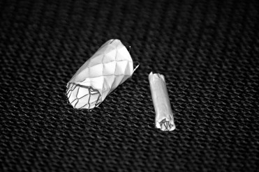
The balloon-expandable covered CP stent consists of platinum iridium wire that is arranged in a “zig” pattern, laser welded at each joint, and over-brazed with gold for added strength ( Figure 1 ). The stent is covered with a sleeve of expanded polytetrafluoroethylene. These stents can be balloon expanded to 12 to 24 mm in diameter. Available lengths range from 16 to 45 mm. The covered CP stent inventory was supplied to participating COAST centers premounted on BIB balloons (NuMed, Inc.) ranging from 12 to 22 mm in diameter. Technical aspects of cardiac catheterization were at the discretion of individual operators and without a standardized protocol. All catheterizations were performed under general anesthesia in a cardiac catheterization laboratory. A percutaneous transfemoral arterial approach was used in all cases. Administration of heparin was not standard because of the burden of noncardiac traumatic injuries and associated risk of hemorrhagic sequelae. After acquisition of hemodynamic data, biplane aortography was performed. Measurements of the aorta and aortic wall injury (aneurysm) were recorded. From these data an adequately sized premounted covered CP stent (typically a diameter 1 to 2 mm larger than the aorta at the level of injury) was chosen for exclusion of TAI. In all cases the covered CP stent was delivered to the site of TAI through a 14Fr-long sheath over a stiff guidewire. Stent implantation occurred with or without the use of rapid right ventricular pacing to attenuate cardiac output and facilitate precise stent positioning during implantation. After postimplantation aortography, the implanted stent was redilated with a larger-diameter balloon in some circumstances. Implantation of a second premounted covered CP stent was performed in cases where significant aortic wall injury persisted after implantation of the first covered stent.

Results
During the COAST protocol 6 covered CP stents were implanted in 4 patients on an emergency-use basis to treat TAI after blunt chest wall trauma. Patient characteristics are presented in Table 1 . All patients were children. A percutaneous approach to repair of TAI was chosen in all cases because of the concomitant presence of severe multiorgan system injuries identified during the preprocedural evaluation. Multidisciplinary consultation with cardiothoracic, neuro, orthopedic, general, and vascular surgical services, when appropriate, occurred in all cases to collectively determine the preferred approach to repair.
| Patient | Age (years)/Sex | Weight (kg) | Height (cm) | Mode of Injury | Aortic Wall Injury | Additional Injuries | |
|---|---|---|---|---|---|---|---|
| Dissection | Aneurysm ⁎ | ||||||
| 1 | 14/M | 50 | 160 | MVA | + | + | TBI, facial fx, lung injuries, pelvic and leg fx |
| 2 | 13/M | 130 | 163 | MVA | + | + | Sternal fx, rib fx, lung injuries, hip, leg, and ankle fx |
| 3 | 14/F | 66 | 174 | MVA | + | + | TBI, facial and skull fx, ABD organ and lung injuries, rib and leg fx |
| 4 | 11/M | 40 | 152 | Boating accident | + | + | TBI, SAH, facial fx, C2–C3 AL, RH, ABD injury, leg fx |
Deployment of the balloon-expandable covered CP stent was technically successful in all attempts. Angiographic images from 2 subjects are displayed in Figures 2 and 3 . Table 2 presents key procedural characteristics. Median fluoroscopy time was 13.2 minutes and median procedure time was 120 minutes. Heparin was administered systemically in 1 case only. Rapid right ventricular pacing was used during stent implantation in 2 cases. In 2 patients a second covered CP stent was deployed in overlapping fashion to completely exclude underlying aortic wall injury. Postimplantation balloon dilation of covered CP stents to a diameter larger than the implantation diameter occurred in 1 case to improve stent–vessel wall apposition owing to a small residual communication with the aneurysm at the stent edge. A tiny residual communication persisted at the completion of this case but was no longer present by CT scan 1 day later. Complete angiographic exclusion of aortic wall injury was achieved in the other 3 cases. In no cases were the origins of brachiocephalic vessels impacted in any way and there was no aortic obstruction after stent implantation.
| Patient | AAo (mm) | TAo (mm) | Isthmus (mm) | DAo (mm) | Size of Stent(s) (mm) | Postimplantation Dilation | Residual Leak ⁎ | Fluoroscopy Time (min) | Procedure Time (min) |
|---|---|---|---|---|---|---|---|---|---|
| 1 | 20.1 | 19 | 19 | — | 20 × 39, 20 × 28 | + (22 mm) | + (trivial) | 18 | 201 |
| 2 | 20.4 | 16.9 | 15.3 | 15.8 | 20 × 45, 20 × 34 | 0 | 0 | 16.1 | 144 |
| 3 | 22.5 | 17.9 | 17.4 | 14.9 | 18 × 39, 18 × 39 † | 0 | 0 | 9.6 | 55 |
| 4 | 18.9 | 12.7 | 13.2 | — | 16 × 39 | 0 | 0 | 10.2 | 96 |
Stay updated, free articles. Join our Telegram channel

Full access? Get Clinical Tree


