Fig. 4.1
Histology of a normal pericardium. Two photomicrographs of the visceral pericardium. (a) Next to the outermost layer of the myocardium. (b) A higher magnification, showing a mesothelial layer that coats loose connective and adipose tissues within which blood vessels and nerves are embedded
Epicardial adipose tissue is more than a fat depot since it is metabolically active and, given its intimate contact with the myocardium (there is no fascia between them), it has been recently associated with the normal physiology and structure of the coronary circulation. Epicardial fat can locally interact with and modulate the coronary arteries through an equilibrium between the paracrine or vasocrine secretion of pericardial inflammatory adipokines on the one hand (eg, tumor necrosis factor alpha, monocyte chemoattractant chemokines, interleukin-6, etc.) and, on the other hand with anti-inflammatory and anti-atherogenic adipokines such as adrenomoduline and adiponectin. Epicardial adipose tissue can play an important role in the inflammatory process at the atheroma plaque. When epicardial fat increases, it extends towards the anterior surface of the heart, and can eventually cover the entire heart with a layer of up to 2 cm in thickness (Fig. 4.2). The increase in epicardial fat in hemodynamic or metabolic pathologies may produce adverse lipotoxic, prothrombotic or proinflammatory effects.
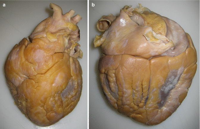
Fig. 4.2
Increase of the epicardial adipose tissue. Anterior (a) and posterior (b) views of a 516 g heart with a substantial increase of the epicardial fat, from a 52-year-old male who died suddenly at home. Autopsy revealed severe coronary atherosclerosis
4.2 Congenital Pathology
Congenital pathology of the pericardium is infrequent and includes complete or partial pericardial absence (agenesis) and congenital cysts.
4.2.1 Pericardial Congenital Defects
Pericardial congenital defects are observed at a frequency of 1/10.000 in autopsies. Complete or bilateral pericardial agenesis is the least frequent form and, in the absence of other associated congenital lesions, is generally asymptomatic. The main clinical manifestation is a mild nonspecific chest pain, likely caused by torsion of the great vessels due to excessive mobility of the heart. Among the partial defects, absence of the left pericardium is the most frequent (Fig. 4.3). Complications can occur when a portion of the myocardium herniates or strangulates at the level of the defect, which may compress the left coronary artery. Clinical symptoms include dyspnea, chest pain, syncope and/or sudden death. Moreover, these defects allow a greater mobility of the heart, which may increase the risk of suffering an aortic dissection after trauma.
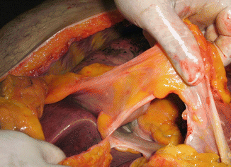
Fig. 4.3
Partial pericardial agenesis. The absence of the inferior pericardium is usually associated with a lack of the left hemidiaphragm, which implies a direct communication between the heart and the abdominal viscera (Courtesy of Dr. Iván Pávez. Medico-Legal Service, Santiago de Chile)
4.2.2 Congenital Pericardial Cysts
Cysts can be uni- or multilocular with a diameter ranging from 1 to 5 cm. Patients are asymptomatic and the cysts are accidentally discovered during thoracic radiologic examinations (a radiodense homogeneous structure), or during autopsy. A differential diagnosis with acquired inflammatory pseudocysts and hydatid cysts should be performed.
Hydatid cysts in the pericardial cavity are generally asymptomatic but they can cause pseudoangina symptoms. Also, if the cysts rupture and spill their contents into the pericardial cavity, they can produce pericardial effusion and cardiac tamponade.
Remarks |
Pericardial pathology very rarely causes sudden cardiac death (SCD) apart from cardiac tamponade, although some cases of sudden death have been described associated with partial pericardial agenesis. |
4.3 Pericardial Effusion. Cardiac Tamponade. Hemopericardium
Under particular circumstances, the fibrous or parietal pericardium distends due to liquid accumulation or pericardial effusion of diverse nature (exudate, transudate, blood). The clinical manifestations will depend on speed at which the fluid accumulates. Thus, effusions of no more than 200–300 cc, if the accumulation is fast (acute effusion), can lead to cardiac tamponade. In contrast, if the effusion is gradual (subacute/chronic effusion), up to 2 l of fluid can accumulate without significantly changing the intrapericardial pressure.
Cardiac tamponade is defined as an accumulation of pericardial fluid that produces an increase in the intrapericardial pressure, which in turn impedes ventricular filling during diastole and reduces the systolic volume. The most frequent pericardial causes of cardiac tamponade are metastatic carcinoma and idiopathic pericarditis. When a distended cardiac sac is found at autopsy the volume of fluid it contains should be determined, and its characteristics evaluated.
Hemopericardium refers to blood in the pericardial cavity, produced typically by a rupture in the myocardium, aortic wall or coronary artery, or due to alterations in the blood coagulation system. From a practical point of view, to correctly assess the dimensions of the blood clot it is convenient to measure the volume of blood and/or to weigh the clot contained within the pericardial cavity.
Serous effusions are most common in patients with cardiac insufficiency and hypoproteinemia, whereas bloody effusion most commonly results from ischaemic or traumatic cardiomyopathy, neoplasms or uraemic or infectious pericarditis. Chylous effusions are extremely rare and result from injury or obstruction of the thoracic duct (Fig. 4.4). Cardiac tamponade secondary to hemopericardium is typically caused by myocardial rupture due to ischaemia or trauma, and by aortic dissection (Fig. 4.5).
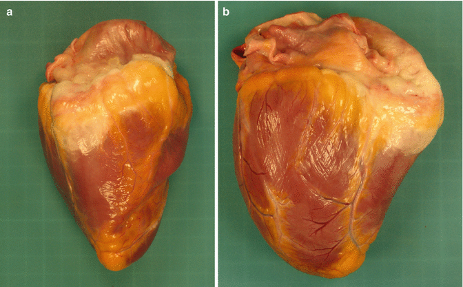
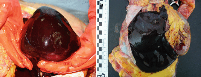
Fig. 4.4
Chylopericardium. Epicardial imbibition from the lympha as a result of chylopericardium due to an iatrogenic tear of the lymphatic thoracic duct. A 21-year-old male who was involved in a car accident and died from head injuries. (a) Right lateral view of the heart. (b) Posterior view of the heart
Fig. 4.5
Cardiac tamponade due to hemopericardium. (a) As the heart is removed, the blood clot adopts the shape of the pericardial sac (Courtesy of Dr. Juan Pedro Hernández Rincón. Institute of Legal Medicine, Murcia). (b) Sudden death by aortic dissection. An incision in the pericardial sac shows the pericardial cavity occupied by a large blood clot that displaces the heart
The pathological features of pericardial effusion are summarised in Table 4.1.
Table 4.1
Pathology of pericardial effusion
In chronic pericardial effusions there is a pericardial fibrous thickening with light or moderate inflammatory infiltration, mainly lymphocytic. |
Subacute pathology responsible for the bleeding, including intramural haematoma on the aortic wall, can be observed in hemopericardium secondary to aortic dissection. |
In myocardial rupture after acute myocardial infarction, the transmural extent of necrosis in the ischaemic myocardium must always be confirmed. < div class='tao-gold-member'>
Only gold members can continue reading. Log In or Register to continue
Stay updated, free articles. Join our Telegram channel
Full access? Get Clinical Tree
 Get Clinical Tree app for offline access

|