Paravalvular Leak
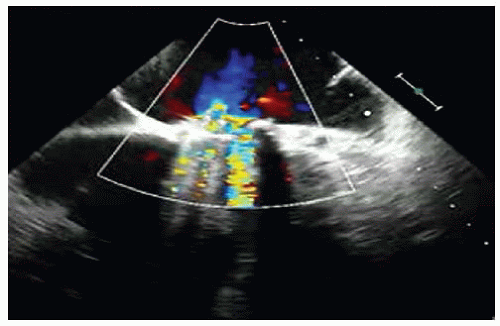
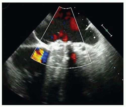
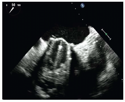
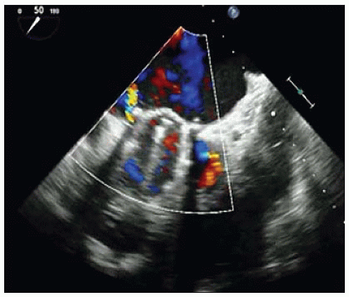
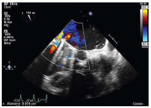
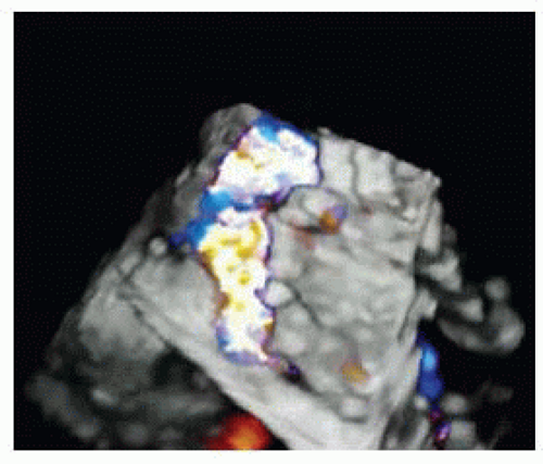
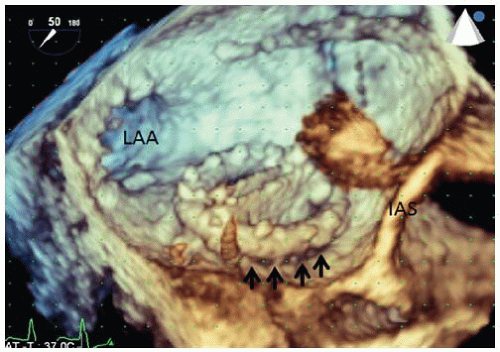
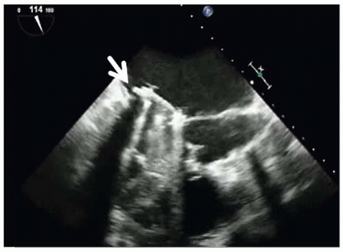
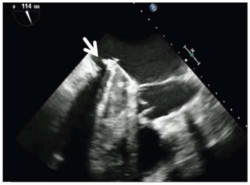
A 57-year-old man had mechanical mitral valve replacement 3 years ago due to severe mitral regurgitation after endocarditis. Six months later, a significant paravalvular leak was verified, and a second operation was performed (St. Jude Medical mechanical valve). In the following months, he developed increasing dyspnea and leg edema. The transesophageal echocardiogram (TEE) study is shown in Videos 39-1 to 39-4 and Figures 39-1, 39-2, 39-3, 39-4, 39-5, 39-6 and 39-7.
QUESTION 1. Which is the correct diagnosis?
A. Paravalvular leak in anterolateral location
B. Paravalvular leak in anteromedial location
C. Paravalvular leak in posteromedial location
D. Paravalvular leak in posterolateral location
View Answer


ANSWER 1: C. One potential complication after cardiac valve replacement is the development of a paravalvular leak, representing one of the most frequent causes for redo surgery.1
Stay updated, free articles. Join our Telegram channel

Full access? Get Clinical Tree


