Overview of Upper Extremity Artery Disease


Overview of Upper Extremity Artery Disease


Steven M. Dean


Although significantly less common than typical atherosclerotic peripheral artery disease (PAD) of the lower extremities, upper extremity artery disease (UEAD) is an important and often challenging cause of arm ischemia and/or distal vasospasm. Hemodynamically significant atherosclerosis is approximately 20 times less common in the arms than in the legs and is usually limited to the proximal or “inflow” arteries. However, vasospastic disorders are more likely to affect the upper than the lower extremities. A variety of unique nonatherosclerotic etiologies can provoke UEAD. Some of these causes are exclusive to the upper limb and include thoracic outlet syndrome (TOS) and occupation-related occlusive disease.

Pathophysiologically, UEAD can be divided into large-artery (proximal to the wrist) and small-artery (distal to the wrist) diseases. A classification scheme for UEAD is outlined in Table 16.1. During the initial patient assessment, the clinician attempts to assign the affected patient into one of these three broad categories. However, prompt and definitive differentiation of these etiologies based on an isolated history and physical is not always possible. Consequently, adjunctive serology testing and noninvasive and/or invasive vascular studies are frequently required to explicitly define the locus and etiology of a patient’s UEAD. Classification overlap may occur between categories. For instance, a patient with thromboangiitis obliterans can be categorized as having both large- and small-artery occlusive disease as well as secondary Raynaud’s syndrome.


ANATOMY AND PHYSIOLOGY OF UPPER EXTREMITY CIRCULATION


Anatomy

The inflow arteries of the upper extremities comprise the innominate and subclavian arteries. Figure 16.1 illustrates the remaining intrinsic vessels of the upper extremity including the axillary, brachial, radial, ulnar, palmar, and digital arteries. Upon traversing the wrist, the radial and ulnar arteries form the deep and superficial palmar arches, respectively (Fig. 16.2). The deep palmar arch yields three palmar metacarpal arteries that anastomose with four common palmar digital branches of the superficial palmar arch. At the level of the finger webs, the common digital arteries bifurcate into paired medial- and lateral-based proper digital arteries that ultimately fuse at the distal tuft (Fig. 16.3).

The radial, ulnar, and proper digital arteries form a parallel arterial system that can preserve normal resting perfusion despite underlying obstructive disease. Consequently, one must remain cognizant that these

duplicate arteries can potentially decrease the sensitivity of noninvasive testing (pressure measurements and plethysmographic waveforms) in identifying UEAD. For example, normal forearm and digital pressures as well as pulsed volume recording (PVR) waveforms may be obtained in the setting of an occluded ulnar artery.


TABLE 16.1 CLASSIFICATION SCHEME FOR UPPER EXTREMITY ARTERIAL DISEASE


I. Thromboembolic Diseases


A. Embolism


– Proximal primary aneurysm


– Post-stenotic aneurysm


– Cardiogenic (including patent foramen ovale)


– Ulcerated aortic arch and/or innominate, subclavian, axillary arteries


B. Thrombosis


– Thrombophilia


  • Antiphospholipid antibody syndrome


  • Heparin-induced thrombocytopenia


  • Proteins S and C, and antithrombin deficiencies; homocysteinemia, factor V Leiden mutation, prothrombin gene mutation (these causes are rare)


– Hematologic disorders


  • Myeloproliferative syndromes, leukemias, hypereosinophilic syndromes


  • Multiple myeloma, Waldenstrom’s macroglobulinemia, cryoglobulinemia


  • Paroxysmal nocturnal hemoglobinuria


– Malignancy


– Vasculitis


II. Arterial Occlusive Diseases


A. Large Artery (proximal to wrist)


– Atherosclerosis


– Large vessel vasculitis (giant cell, Takayasu’s)


– Medium vessel vasculitis (polyarteritis nodosa, Kawasaki’s disease)


– Thromboangiitis obliterans


– Fibromuscular dysplasia


– Radiation fibrosis


– Entrapment (TOS, axillary artery crutch injury)


– Aneurysms (subclavian through radioulnar level)


B. Small Artery (distal to wrist)


– Autoimmune diseases


– Small- and medium-sized vasculitis


– Thromboangiitis obliterans


– Hypothenar hammer syndrome, thenar hammer syndrome, hand-arm vibration syndrome


– Hematologic disorders (as above)


– Thrombophilia (as above)


– Malignancy


– Frostbite


– Calciphylaxis/calcific azotemic arteriopathy


III. Vasospastic Diseases


A. Large Artery


– Ergotamines


– Methamphetamine and related medications


– Cocaine


B. Small Artery


– Primary Raynaud’s syndrome


– Secondary Raynaud’s syndrome


Anomalous Circulation

In order to avoid a false-positive diagnosis of UEAD, a clinician should be aware that a variety of arterial anomalies may occur in the upper extremity. Arterial variants of the upper extremity are listed in Table 16.2.


Collateral Pathways

In addition to the previously described dual arterial system, the upper extremity possesses an abundance of complementary arterial tributaries that can function as collaterals in the presence of significant UEAD. This highly redundant system can significantly delay the onset of ischemic symptoms despite underlying arterial occlusive disease. The prototypical upper extremity collateral pathway is exemplified by the vertebral artery functioning as a collateral conduit in the setting of a proximal subclavian (or innominate) artery severe stenosis or occlusion. Figure 16.1 illustrates several arterial branches that can function as collaterals in the event of a brachial artery occlusion (e.g., profunda brachii, radial recurrent, superior and inferior ulnar collaterals).


Physiologic and Pathophysiologic Vasospasm

The sympathetic nervous system regulates local perfusion via adrenergic receptors on the smooth muscle of cutaneous vessels. Typically, the cutaneous circulation assumes a predominant role in preserving a stable core body temperature by adjusting the surface blood flow contingent on the ambient temperature. For example, cold temperatures evoke shunting of blood away from the surface, consistent with physiological or “normal” vasospasm. Conversely, exposure to warm environments augments perfusion to the skin. Raynaud’s syndrome arises when this normal or physiological response to ambient cold temperatures is exaggerated and


digital perfusion intermittently ceases. Physiological vasospasm is never associated with complete cessation of digital perfusion. Primary Raynaud’s syndrome only involves functional alterations, whereas secondary Raynaud’s syndrome also involves associated structural microvascular abnormalities. The pathogenesis of Raynaud’s syndrome includes defects in one of more of these three systems: (a) vascular (endothelial dysfunction; structural defects), (b) neural (central dysfunction; impaired vasodilation; impaired vasoconstriction), and (c) intravascular abnormality (increased platelet activation and aggregation; impaired fibrinolysis).


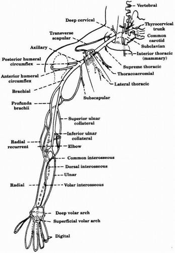
FIGURE 16-1. Arterial supply of the upper extremity.


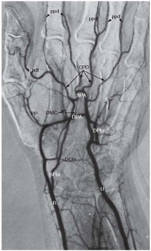
FIGURE 16-2. Normal arteriogram of the hand showing the arches (superficial and deep). (CPD, common palmar digital; DCbr, dorsal carpal branch; DMC, dorsal metacarpal; DPA, deep palmar arch; DPbr, deep palmar branch of ulnar; PP, princeps pollicis (main artery to the thumb); ppd, palmar proper digital; ppdt, palmar proper digital thumb; R, radial; SPA, superficial palmar arch; SPbr, superficial palmar branch of radial; U, ulnar.) (Adapted from Kadir S. Atlas of normal and variant angiographic anatomy. W. B. Saunders Company, 1991.)


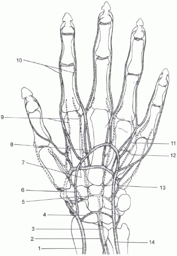
FIGURE 16-3. Arterial anatomy of the hand. (1, Radial; 2, Anterior interosseus; 3, Posterior interosseus; 4, Palmar carpal branch; 5, Dorsal carpal branch forming the dorsal carpal rete; 6, Superficial palmar branch of the radial artery; 7, Deep palmar arch; 8, Superficial palmar arch; 9, Dorsal metacarpal arteries; 10, Proper palmar digital arteries; 11, Common palmar digital arteries; 12, Palmar metacarpal; 13, Deep palmar branch of the ulnar artery; 14, Ulnar artery.) (Adapted from Kadir S. Atlas of normal and variant angiographic anatomy. W. B. Saunders Company, 1991.)


TABLE 16.2 ARTERIAL VARIANTS OF THE UPPER EXTREMITY


Variant


Frequency of Occurrence in the Population (%)


Aortic arch and great vessels


Common origin of the right brachiocephalic and left common carotid arteries (bovine arch)


22


Left vertebral artery origin directly from the aorta


4-6


Brachial artery


Radial artery origin from the brachial or axillary


15-20


Ulnar artery origin from the brachial or axillary


1-3


Accessory (duplicated) brachial artery (rejoin in the antecubital fossa)


0.1-0.2


Radial artery


Radial artery origin from the brachial or axillary


15-20


Aplasia, hypoplasia, and duplication


<1


Ulnar artery

Only gold members can continue reading. Log In or Register to continue

Stay updated, free articles. Join our Telegram channel

Jun 12, 2016 | Posted by in CARDIOLOGY | Comments Off on Overview of Upper Extremity Artery Disease

Full access? Get Clinical Tree

Get Clinical Tree app for offline access